Nickel Concentrations in Serum of Patients with Acute Myocardial Infarction or Unstable Angina Pectoris.

Abstract

Nickel was measured, by electrothermal atomic absorption spectrophotometry, in sera from (a) 30 healthy adults, (b) 54 patients with acute myocardial infarction, (c) 33 patients with unstable angina pectoris without infarction, and (d) five patients with coronary atherosclerosis who developed cardiac ischemia during treadmill exercise. Mean (and SD) concentrations in Group a were 0.3 (0.3) microgram/L (range less than 0.05-1.1 microgram/L). Within 72 h after hospital admission, hypernickelemia (Ni greater than or equal to 1.2 microgrheart-1414885-639x650am/L) was found in 41 patients of group b (76%) and in 16 patients of group c (48%). Hypernickelemia was found before and after exercise in one patient of Group d (20%). Peak values averaged 3.0 micrograms/L (range 0.4-21 micrograms/L) in Group b, 1.5 microgram/L (range less than 0.05-3.3 micrograms/L) in Group c. In Group b, the mean time interval between the peak values for creatine kinase activity and for nickel was 18 h. Serum nickel concentrations were unrelated to age, sex, time of day, cigarette smoking, medications, clinical complications, or outcome. Mechanisms and sources of release of nickel into the serum of patients with acute myocardial infarction or unstable angina pectoris are conjectural, but hypernickelemia may be related to the pathogenesis of ischemic myocardial injury.

www.clinchem.org/content/31/4/556.full.pdf

Blood lead below 0.48 micromol/L (10 microg/dL) and mortality among US adults.

Menke A1, Muntner P, Batuman V, Silbergeld EK, Guallar E.

Abstract

Background— Blood lead levels above 0.48 μmol/L (10 μg/dL) in adults have been associated with increased risk of cardiovascular, cancer, and all-cause mortality. The objective of the present study was to determine the association between blood lead levels below 0.48 μmol/L and mortality in the general US population.

Methods and Results— Blood leadlead-1565598-640x480 levels were measured in a nationally representative sample of 13 946 adult participants of the Third National Health and Nutrition Examination Survey recruited in 1988 to 1994 and followed up for up to 12 years for all-cause and cause-specific mortality. The geometric mean blood lead level in study participants was 0.12 μmol/L (2.58 μg/dL). After multivariate adjustment, the hazard ratios (95% CI) for comparisons of participants in the highest tertile of blood lead (≥0.17 μmol/L [≥3.62 μg/dL]) with those in the lowest tertile (<0.09 μmol/L [<1.94 μg/dL]) were 1.25 (1.04 to 1.51; Ptrend across tertiles=0.002) for all-cause mortality and 1.55 (1.08 to 2.24; Ptrend across tertiles=0.003) for cardiovascular mortality. Blood lead level was significantly associated with both myocardial infarction and stroke mortality, and the association was evident at levels >0.10 μmol/L (≥2 μg/dL). There was no association between blood lead and cancer mortality in this range of exposure.

Conclusions— The association between blood lead levels and increased all-cause and cardiovascular mortality was observed at substantially lower blood lead levels than previously reported. Despite the marked decrease in blood lead levels over the past 3 decades, environmental lead exposures remain a significant determinant of cardiovascular mortality in the general population, constituting a major public health problem.

http://circ.ahajournals.org/content/114/13/1388.long

Depression of cholesterol levels in human plasma following ethylenediamine tetracetate and hydralazine

H. Mitchell Perry Jr., M.D.
Henry A. Schroeder, M.D.
blood-test-1545982-639x493

Abstract

Twenty-two patients received from 3 to 27 Gm. of intravenous EDTA. Six times after an average total dose of 18 Gm., and without change in other metal binding drugs, the total fasting plasma cholesterol fell an average of 75 mg. per 100 ml. of plasma. Initially low values were altered less. The average depression in 112 unselected hypertensive patients was 29 mg. following oral hydralazine therapy; however, among the 66 with high control levels the fall was 48.2 mg. in the first six months, and thereafter 43.5 mg. per 100 ml. of plasma. In 10 hypertensive patients who received an average of 13 Gm. of EDTA the blood pressure decreased an equivocal Math Eq. Hg, although for 6 who took autonomic-blocking agents there was a simultaneous reduction of 34 per cent in the requirement for that drug. Of 9 patients who received single doses of 4 Gm. or more of EDTA, 8 quickly developed fever, and in 2 who received large total doses in relation to their body weight, mucocutaneous lesions were observed.

Full PDF available for purchase: http://www.jclinepi.com/article/0021-9681%2855%2990151-X/abstract

Disappearance of immune deposits with EDTA chelation therapy in a case of IgA nephropathy

by Lin JL, Lim PS

Abstract

In this report, we describe the development of renal function impairment in a 33-year-old patient with mesangial IgA nephropathy and a history of recent gout. Increased body lead burden was identified with a positive EDTA mobilization test. The patient was treated with 1 g of edetate disodium calcium weekly for 2 months until normalization of urinary lead excretion. Improvement of renal function and proteinuria were noted. It was even more interesting to find that both immunofluorescence and electron microscopy studies of the second bioarteriography-1562063-640x600psy specimen revealed the loss of previous mesangial immune deposits. Our case demonstrated that lead may be a nonspecifically damaging factor related to the deterioration of renal function in patients with preexisting renal disease. Moreover, the disappearance of mesangial immune deposits after chelation therapy has not been previously documented. The pathogenetic basis of this observation is unknown, and its causal relationship with lead requires further elucidation.

http://www.ncbi.nlm.nih.gov/pubmed/1292346


The Scientific Basis for EDTA Chelation Therapy

In the 1980’s, chelation therapy demonstrated great success in treating chronic circulatory problems in several nations around the world. 30 years later, chelation therapy remains a controversial practice that is largely misunderstood in the mainstream population.

To help inform both the public and other physicians of the benefits of chelation therapy, Edward W. Macdonagh collected research articles containing several studies done globally on chelation. The research was collected and put into a book titled “The Scientific Basis of EDTA Chelation Therapy” which was published in 1981.

Below is the table of contents for the collection of articles by Edward W. Macdonagh and Charles Rudolph DO. Abstracts are available at http://www.mcdonaghmed.com/chelation-therapy/

McDonagh Medical Center Research Articles

  • Serum Cholesterol and the Aging Process
  • The Relationship of Subtle Differences in Fasting Blood Glucose with Subtle Difference in the Electrocardiogram: A Study of the PR Interval
  • The Homeostatic Effect of EDTA with Supportive Multivitamin Trace Mineral Supplementation upon High-Density Lipoproteins (HDL)
  • The Effect of Intravenous Disodium Ethylenediaminetetraacetic Acid (EDTA) upon Blood Cholesterol in a Private Practice Environment
  • An Oculocerebrovasculometic Analysis of the Improvements in Arterial Stenosis Following EDTA Chelation Therapy
  • The Effect of EDTA Chelation Therapy Plus Supportive Multivitamin-Trace Mineral Supplementation upon Renal Function: A Study in Serum Creatinine
  • The Glycohemoglobin (HGBA1C) Distribution in EDTA-Chelation-Eligible Patients
  • The Influence of EDTA Salts Plus Multivitamin-Trace Mineral Therapy Upon Total Serum Cholesterol/High Density Lipoprotein Cholesterol
  • The Chelation Carrier Solution: An Analysis of Osmolarity and Sodium Content
  • The Nutrition-Prevention Connection
  • The “Health” of the Parolee: Clinical Considerations
  • The Effect of EDTA Chelation Therapy with Multivitamin/Trace Mineral Supplementation Upon Reported Fatigue
  • Effect of EDTA Chelation and Supportive Multivitamin/Trace Mineral Supplementation With and Without Physical Activity on the Heat Rate
  • The Effect of ETDA Chelation Therapy Plus Multivitamin/Trace Mineral Supplementation Upon Vascular-Dynamics (Ankle/Brachial Systolic Blood Pressure)
  • The “Clinical Change” in Patients Treated with EDTA Chelation Plus Multivitamin/Trace Mineral Supplementation
  • The Psychotherapeutic Potential of EDTA Chelation
  • Effect of EDTA Chelation and Supportive Multivitamin/Trace Mineral Supplementation With and Without Physical Activity Upon Systolic Blood Pressure
  • The Effect of EDTA Chelation Therapy Plus Supportive Multivitamin/Trace Mineral Supplementation Upon Renal Function: A Study in Blood Urea Nitrogen
  • The Effect of Intravenous Disodium Ethlenediaminetetraacetic Acid (EDTA) Plus Supportive Multivitamin/Trace Mineral Supplementation Upon Fasting Serum Calcium
  • Trace Element Patterning in Degenerative Diseases
  • The Effect of Intravenous Disodium Ethlenediaminetetraacetic Acid Upon Bone Density Levels
  • Effect of EDTA Chelation and Supportive Multivitamin / Trace Mineral Supplementation on Chronic Lung Disorders: A Study of FVC and FEV
  • Effect of EDTA Chelation and Supportive Multivitamin/Trace Mineral Supplementation on Carotid Circulation: Case Report
  • Effect of EDTA Chelation Therapy on Aortic Calcium in Rabbits on Atherogenic Diets; Quantitative and Histochemical Studies
  • An Observation of the Effect of EDTA Chelation and Supportive Multivitamin/Trace Mineral Supplementation on Blood Platelet Volume: A Brief Communication
  • Effect of EDTA Chelation on Serum Iron
  • A Nonsurgical Approach to Obstruction Carotid Stenosis Using EDTA Chelation
  • Effects of EDTA Chelation on Chronic Degenerative Diseases
  • Magnetic Resonance Imaging Evidence of a Reduction in Disc Herniation Using a Combination of EDTA Chelation and Joint Reconstructive Therapy
  • Noninvasive Treatment for Sequellae of Failed Coronary Blood Circulation; 100% Occlusion of Left Anterior Descending Coronary Artery, 30% Stenosis Right Coronary Artery, and Left Ventricular Contractility Deficit
  • Visual Field Evidence of Macular Degeneration Reversal Using a Combination of EDTA Chelation and Multiple Vitamin and Trace Mineral Therapy
  • Renal Artery Stenosis Reversal in a Hypertensive Individual, Using a Combination of EDTA Chelation and Multiple Vitamin and Trace Mineral Therapy


EDTA Redistribution of Lead and Cadmium Into the Soft Tissues in a Human With a High Lead Burden – Should DMSA Always Be Used to Follow EDTA in Such Cases?

by Crinnion WJ

anatomy-1428238-639x609Abstract

Intravenous sodium calcium ethylene diamine tetra acetic acid (EDTA) and oral 2,3-dimercaptosuccinic acid (DMSA) have both been used to reduce the burden of lead in humans. Each of these agents enhances the mobilization of lead from different areas of the body – EDTA from the trabecular bone and DMSA from the soft tissue. A study of Korean battery workers revealed that EDTA appeared to increase the soft tissue burden of lead, resulting in increased levels of aminolevulinic acid and greater subsequent lead mobilization with DMSA. This case report discusses a patient with a higher-than-normal lead burden who exhibited increased tissue lead burden after intravenous EDTA. The elevated tissue burden of lead was still present, albeit lower, after five consecutive days of oral DMSA therapy. If this single case is representative of a typical human response to the use of intravenous (IV) EDTA for lead, then it suggests that all persons undergoing such treatment should be administered oral DMSA for a minimum of one week after EDTA treatment.

 www.altmedrev.com/publications/16/2/109.pdf

TACT Trial: EDTA Chelation Therapy for Atherosclerosis

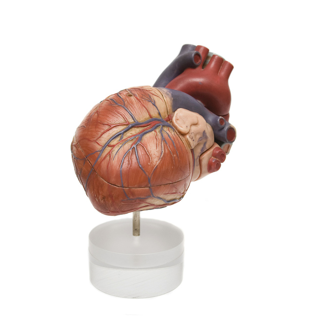
Effect of Disodium EDTA Chelation Regimen on Cardiovascular Events in Patients With Previous Myocardial Infarction

Lamas-Gervasio-CARDIOLOGYGervasio A. Lamas, MD; Christine Goertz, DC, PhD; Robin Boineau, MD, MA; Daniel B. Mark, MD, MPH; Theodore Rozema, MD; Richard L. Nahin, PhD, MPH; Lauren Lindblad, MS; Eldrin F. Lewis, MD, MPH; Jeanne Drisko, MD; Kerry L. Lee, PhD ;

Importance Chelation therapy with disodium EDTA has been used for more than 50 years to treat atherosclerosis without proof of efficacy.

Objective To determine if an EDTA-based chelation regimen reduces cardiovascular events.

Design, Setting, and Participants Double-blind, placebo-controlled, 2 × 2 factorial randomized trial enrolling 1708 patients aged 50 years or older who had experienced a myocardial infarction (MI) at least 6 weeks prior and had serum creatinine levels of 2.0 mg/dL or less. Participants were recruited at 134 US and Canadian sites. Enrollment began in September 2003 and follow-up took place until October 2011 (median, 55 months). Two hundred eighty-nine patients (17% of total; n=115 in the EDTA group and n=174 in the placebo group) withdrew consent during the trial.

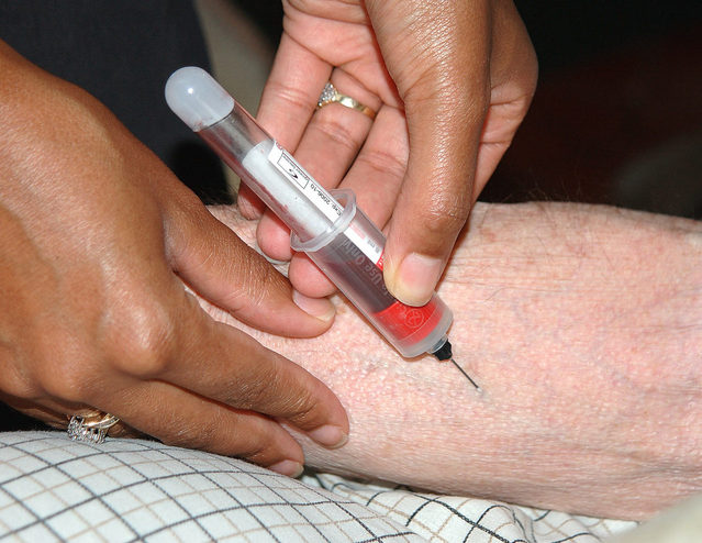
Interventions Patients were randomized to receive 40 infusions of a 500-mL chelation solution (3 g of disodium EDTA, 7 g of ascorbate, B vitamins, electrolytes, procaine, and heparin) (n=839) vs placebo (n=869) and an oral vitamin-mineral regimen vs an oral placebo. Infusions were administered weekly for 30 weeks, followed by 10 infusions 2 to 8 weeks apart. Fifteen percent discontinued infusions (n=38 [16%] in the chelation group and n=41 [15%] in the placebo group) because of adverse events.

Main Outcome Measures The prespecified primary end point was a composite of total mortality, recurrent MI, stroke, coronary revascularization, or hospitalization for angina. This report describes the intention-to-treat comparison of EDTA chelation vs placebo. To account for multiple interim analyses, the significance threshold required at the final analysis was P = .036.

Results Qualifying previous MIs occurred a median of 4.6 years before enrollment. Median age was 65 years, 18% were female, 9% were nonwhite, and 31% were diabetic. The primary end point occurred in 222 (26%) of the chelation group and 261 (30%) of the placebo group (hazard ratio [HR], 0.82 [95% CI, 0.69-0.99]; P = .035). There was no effect on total mortality (chelation: 87 deaths [10%]; placebo, 93 deaths [11%]; HR, 0.93 [95% CI, 0.70-1.25]; P = .64), but the study was not powered for this comparison. The effect of EDTA chelation on the components of the primary end point other than death was of similar magnitude as its overall effect (MI: chelation, 6%; placebo, 8%; HR, 0.77 [95% CI, 0.54-1.11]; stroke: chelation, 1.2%; placebo, 1.5%; HR, 0.77 [95% CI, 0.34-1.76]; coronary revascularization: chelation, 15%; placebo, 18%; HR, 0.81 [95% CI, 0.64-1.02]; hospitalization for angina: chelation, 1.6%; placebo, 2.1%; HR, 0.72 [95% CI, 0.35-1.47]). Sensitivity analyses examining the effect of patient dropout and treatment adherence did not alter the results.

Conclusions and Relevance Among stable patients with a history of MI, use of an intravenous chelation regimen with disodium EDTA, compared with placebo, modestly reduced the risk of adverse cardiovascular outcomes, many of which were revascularization procedures. These results provide evidence to guide further research but are not sufficient to support the routine use of chelation therapy for treatment of patients who have had an MI.

Trial Registration clinicaltrials.gov Identifier: NCT00044213

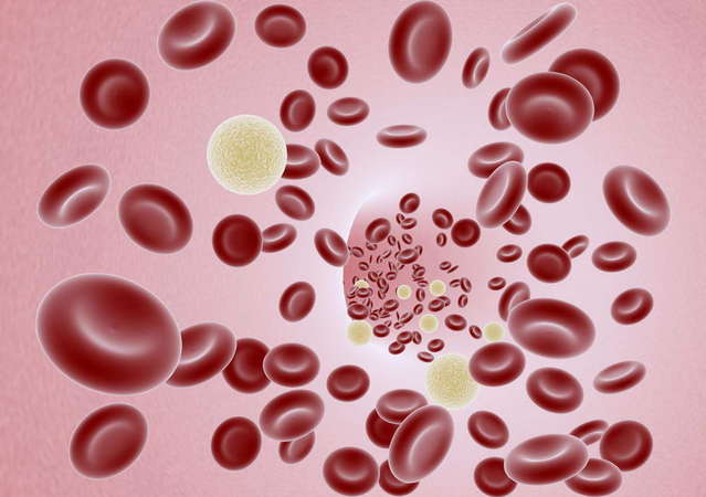
Treatment of lead toxicity with chelation was first reported with EDTA in the early 1950s.1Apparent success in reducing metastatic calcium deposits2 led Clarke et al3 in 1956 to treat angina patients with EDTA, and others to use chelation for various forms of atherosclerotic disease.4– 6Chelation therapy evolved to constitute infusions of vitamins and disodium EDTA, a drug that binds divalent and some trivalent cations, including calcium, magnesium, lead, cadmium, zinc, iron, aluminum, and copper, facilitating their urinary excretion.7,8

Over the next decades, based on favorable anecdotal and case report experience, chelation practitioners increased their use of EDTA for coronary and peripheral artery disease. The 2007 National Health Statistics Report compared chelation use since 2002 and noted an increase of 68%, from 66 000 to 111 000 adults using chelation therapy,9 although the indications for therapy were not clearly defined, and the prevalence of use of chelation therapy for cardiovascular disease is unknown.

Three small clinical trials have assessed the effects of chelation on surrogate outcomes, such as walking distance in patients with claudication (2 trials with 185 patients total) and time to exercise-induced ischemia in patients with coronary disease (1 trial with 84 patients). These studies did not find any evidence of treatment efficacy but were underpowered for evaluation of clinical events.10– 12 As a consequence, mainstream medical organizations consider the therapeutic value of chelation for atherosclerotic vascular disease unproven13 and the use of this therapy potentially dangerous. Disodium EDTA, particularly when infused too rapidly, may cause hypocalcemia and death.14 The Trial to Assess Chelation Therapy (TACT) was conducted to respond to the public health problem posed by EDTA chelation therapy: large numbers of patients being exposed to undefined risks for unproven benefits.

http://jama.jamanetwork.com/article.aspx?articleid=1672238

Chelation Therapy for Peripheral Arterial Occlusive Disease

blood-1237550-639x450E. Ernst, MD, PhD, FRCP (Edin)

Abstract:

Chelation therapy is viewed, promoted, and practiced as a form of complementary/alternative medicine (see References 1 and 21 2 ). It uses repeated intravenous administration of EDTA, usually in combination with vitamins, trace elements, and iron supplements as a treatment for a variety of diseases.3 4 5 Oral chelation therapy also has been advocated (see Reference 66 ) but will be excluded from this review. Among others, chelation therapy is claimed to be effective in reversing the arteriosclerotic disease process,7 8 in particular peripheral arterial occlusive disease (PAOD). Proponents claim that it can be an alternative for amputation in severe cases.8 This goes back to an observation made in the 1950s, when it was noted that patients undergoing EDTA therapy for lead poisoning felt a relief of angina pectoris after this therapy.9 While various authors9 10 11 12 13 1415 have subsequently reported promising results in uncontrolled studies, others have been unable to confirm these in similarly designed trials.16 17 The debate of whether or not it is a useful form of therapy for PAOD therefore continues.18 19 2021 22 23 24 25

Meanwhile, numerous clinics have been set up in Europe, the United States, and elsewhere using and promoting chelation therapy for PAOD. In the light of recent trial data, it is therefore timely to systematically review the evidence from randomized, placebo-controlled, double-blind trials and ask whether chelation therapy for PAOD is safe and effective.

http://circ.ahajournals.org/content/96/3/1031.long


EDTA chelation therapy in the treatment of vascular disease.

Chappell LT1, Janson M.

Abstract

A retrospective analysis of treatment results from 2870 patients, with various chronic degenerative and age-associated diseases, who were treated with di-sodium magnesium EDTA chelation therapy, suggests that the case against EDTA Chelation Therapy should be re-opened.

Using qualitative but never-the-less standardized criteria for improvement, our analysis shows that EDTA Chelation Therapy resulted in “marked” improvement in 76.89% and “good” improvement in 16.56% of patients with ischemic heart disease; also, “marked” improvement in 91% and “good” improvement in 7.6% of patients with peripheral vascular disease and intermittent claudication. In a group of patients with cerebro-vascular and other degenerative cerebral diseases, 24% had “marked” improvement, and 30% had “good” improvement. Of four patients with scleroderma, three had “marked” improvement and one had “good” improvement. Seventy-five percent of all of the patients had “marked” improvement in “geriatric symptomatology of vascular origin”.

The authors recommend renewed study of EDTA Chelation Therapy. The possibility of a “tomato effect”, i.e., a drug which works, but the majority of physicians believe that it doesn’t work, needs to be ruled out. A favorable climate needs to be created, in which FDA-approved studies of its usefulness in treating peripheral vascular disease can take place.

http://www.ncbi.nlm.nih.gov/pubmed/8820322

Acute prooxidant effects of vitamin C in EDTA chelation therapy and long-term antioxidant benefits of therapy.

Hininger I1, Waters ROsman MGarrel CFernholz KRoussel AMAnderson RA.

Chelation therapy is thought to not only remove contaminating metals but also to decrease free radical production. EDTA chelation therapy, containing high doses of vitamin C as an antioxidant, is often used in the treatment of diseases such as diabetes and cardiovascular diseases but the effectiveness of this treatment may be variable and its efficacy has not been demonstrated conclusively. The objective of this work was to determine if the vitamin C added to standard chelation therapy cocktails was prooxidant. We administered a standard EDTA cocktail solution with or without 5 g of sodium ascorbate. One hour following the standard chelation therapy, there were highly significant prooxidant effects on lipids, proteins, and DNA associated with decreased activities of RBC glutathione peroxidase and superoxide dismutase while in the absence of sodium ascorbate, there were no acute signs of oxidative damage. After 16 sessions of standard chelation therapy, the acute prooxidant effects of vitamin C remained, but, even in the absence of nutrient supplements, there were beneficial long-term antioxidant effects of chelation therapy and plasma peroxide levels decreased. In conclusion, multiple vitamins-1471906-640x480sessions of EDTA chelation therapy protect lipids against oxidative damage. However, standard high amounts of vitamin C added to EDTA chelation solutions also display short term prooxidant effects. The added benefits of lower levels of vitamin C in chelation therapy need to be documented.

http://www.ncbi.nlm.nih.gov/pubmed/15917185