Holliday, MD: Carotid Restenosis: A Case for EDTA Chelation

By H. Joseph Holliday, MD, FACA, RVT

hollidayABSTRACT

Carotid restenosis has been found in up to 25% of patients after carotid endarterectomy. The most common cause of restenosis is continuation of the atherosclerotic process. Surgery can be beneficial in stroke prevention and should be considered in those patients at high risk for stroke. However, surgery does not arrest the disease of atherosclerosis. This report demonstrates a 10% reduction in the degree of stenosis in a patient treated with EDTA chelation for restenosis of a carotid artery after endarterectoy. EDTA chelation does arrest and reverse atherosclerosis and should be used in conjuction with surgery or as a primary treatment for carotid restenosis as well as for vascular occlusive disease in any artery whether initial or recurrent.

Click for full text PDF

Effect of chelation therapy on progressive diabetic nephropathy in patients with type 2 diabetes and high-normal body lead burdens.

Chen KH1, Lin JL, Lin-Tan DT, Hsu HH, Hsu CW, Hsu KH, Yen TH., 2012

diabetes-1327517-640x480BACKGROUND:

A previous study in type 2 diabetic patients with high-normal body lead burdens showed that EDTA chelation therapy for 3 months slows progressive diabetic nephropathy during a 12-month follow-up. The effect of a longer course of therapy on kidney function decrease over a longer follow-up is not known.

STUDY DESIGN:

A 12-month run-in phase, then a randomized single-blind study with a 27-month intervention.

SETTING & PARTICIPANTS:

University medical center; 50 patients (serum creatinine, 1.5-3.9 mg/dL) with high-normal body lead burden (≥80-<600 μg) were randomly assigned to the treatment and control groups.

INTERVENTION:

The treatment group received weekly chelation therapy for 3 months to reduce their body lead burden to <60 μg and then as needed for 24 months to maintain this level. The control group received placebo for 3 months and then weekly for 5 weeks at 6-month intervals for 24 months.

OUTCOMES:

The primary end point was change in estimated glomerular filtration rate (eGFR) over time. A secondary end point was a 2-fold increase in baseline serum creatinine level or the requirement for renal replacement therapy.

MEASUREMENTS:

Body lead burdens were assessed by EDTA mobilization tests and eGFR was calculated using the equation for Chinese patients with type 2 diabetes.

RESULTS:

Mean baseline eGFRs in the treatment and control groups were similar. After 3 months of chelation therapy, the change in eGFR in the treatment group (+1.0 ± 4.8 mL/min/1.73 m(2)) differed significantly from that in the control group (-1.5 ± 4.8 mL/min/1.73 m(2); P = 0.04). In the subsequent 24-month intervention, the yearly rate of decrease in eGFR (5.6 ± 5.0 mL/min/1.73 m(2) per year) in the treatment group was slower than that (9.2 ± 3.6 mL/min/1.73 m(2) per year; P = 0.04) in the control group. 17 (68%) control-group patients and 9 (36%) treatment-group patients achieved the secondary end point.

LIMITATIONS:

Small sample size, not double blind.

CONCLUSIONS:

A 27-month course of EDTA chelation therapy retards the progression of diabetic nephropathy in type 2 diabetic patients with high-normal body lead burdens.

http://www.ncbi.nlm.nih.gov/pubmed/22721929

Clinical Benefit of EDTA-based Chelation Therapy and High-dose Oral Multivitamins and Multiminerals in TACT- An Expanded Comparison of 2 Factorial Groups

Lamas, et al. (See below for other contributors)

Background: Chelation therapy, in combination with high dose oral multivitamins and multiminerals (MV), has been used to treat atherosclerotic disease. This analysis of the NIH-funded Trial to Assess Chelation Therapy (TACT) focuses on the comparison of 2 treatment groups chelation + MV versus placebo infusions + placebo MV, evaluating a treatment strategy reflecting clinical practice, and not previously presented in detail.

Methods: TACT, a multi-center, double-blind, placebo-controlled, 2 X 2 factorial trial of EDTA chelation and MV enrolled 1708 patients age ≥ 50 years, MI ≥ 6 weeks prior, creatinine ≤ 2.0, assigned to 40 IV chelation or placebo infusions and a 28-component oral MV or placebo. Primary endpoint was death, MI, stroke, coronary revascularization, or hospitalization for angina. Secondary endpoint was cardiovascular mortality, MI, or stroke. We describe the intent-to-treat comparison of 2 factorial groups: chelation + MV (n=421) vs placebo chelation + placebo MV (n=437). Treatment comparisons were by log rank test.

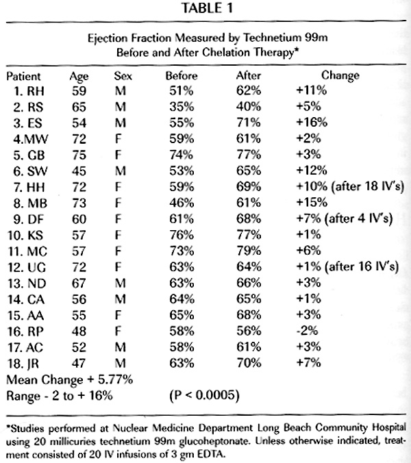
Results: The median age was 65 years, 83% had prior coronary revascularization, and 73% were on statins. Key baseline characteristics were similar between groups. Primary endpoint occurred in 108 (26%) patients in the chelation + MV, and 139 (32%) in the placebo + placebo group (p=0.016). The 5-year Kaplan Meier estimates for the primary endpoint in the chelation + high-dose vitamin group was 31.9%, and in the placebo infusions + placebo vitamin group 40.2%. The secondary endpoint occurred in 39 (9%) of chelation + MV and in 58 (13%) of placebo + placebo (p=0.045). The point estimates for each component of the combined endpoints were all <1.0 and consistent with the overall effect (Table).

Conclusions: In stable patients with a history of MI on appropriate evidence based medical therapy, the beneficial effect of the combination of high-dose vitamins and chelation therapy was statistically significant and of potential clinical relevance.

chelation graph

http://circ.ahajournals.org/cgi/content/meeting_abstract/128/22_MeetingAbstracts/A11054

Gervasio A Lamas1; Richard L Nahin2; Lauren Lindblad3; Christine Goertz4; Robin Boineau5; Terry Chappell6; Greg Flaker7; Theodore Rozema8; Tammy Born9; Mario Stylianou10; Eldrin F Lewis11; Daniel B Mark12; Kerry L Lee3

1 Div of Cardiology, Mount Sinai Med Cntr, Miami Beach, FL
2 31 Cntr Dr, MS 2182, National Cntr for Complementary and Alternative Medicine, Bethesda, MD
3 Biostatistics & Bioinformatics, Duke Clinical Rsch Institute, Durham, NC
4 Chiropractic Rsch, Palmer College of Chiropractic, Davenport, IA
5 HEART FAILURE & ARRHYTHMIAS BRANCH, National Heart, Lung and Blood Institute, Bethesda, MD
6 N/A, Celebration of Heath Association, Bluffton, OH
7 Cardiovascular Medicine, Univ of Missouri Health Care, Columbia, MO
8 N/A, Biogenesis Med Cntr, Landrum, SC
9 N/A, Born Preventive Health Care Clinic, Grand Rapids, MI
10 Biostatistics, National Heart, Lung and Blood Institute, Bethesda, MD
11 Cardiovascular Div, Brigham & Women’s Hosp, Boston, MA
12 Medicine – Cardiology, Duke Clinical Rsch Institute, Durham, NC

EDTA Chelation Therapy: Efficacy in Arteriosclerotic Coronary Heart Disease

H. Richard Casdorph, MD, PhD

Dr. Casdorph is Assistant Clinical Professor of Medicine at the University of California Medical School, Irvine California

ABSTRACT: EightCasdorph_hearteen patients with documented arteriosclerotic heart disease were studied utilizing the radioactive isotope technetium 99m to measure left ventricular ejection fraction before and after the administration of EDTA chelation therapy. A statistically significant improvement in left ventricular ejection fraction occurred in this group of patients. The patients were tested at rest, but conditions under which tests were performed were controlled to be identical before and after chelation.

http://drcranton.com/chelation/casdor1.htm


Chelation therapy for patients with elevated body lead burden and progressive renal insufficiency. A randomized, controlled trial.

Chelation therapy for patients with elevated body lead burden and progressive renal insufficiency. A randomized, controlled trial.

Lin JL1, Ho HH, Yu CC, 1999

lab-work-1575843-640x960Abstract

BACKGROUND:

Nephropathy is known to occur in persons exposed to high levels of lead, but the question of whether long-term exposure to low levels of environmental lead is associated with impaired renal function remains controversial.

OBJECTIVE:

To examine whether chelation therapy slows the progression of renal insufficiency in patients with mildly elevated body lead burden.

DESIGN:

Randomized, controlled trial.

SETTING:

Academic medical center in Taiwan.

PATIENTS:

32 patients with chronic renal insufficiency (serum creatinine level > 132.6 micromol/L [1.5 mg/dL] and < 353.8 micromol/L [4.0 mg/dL]), mildly elevated body lead burden (> 0.72 micromol [150 microg] of lead per 72-hour urine collection and < 2.90 micromol [600 microg] of lead per 72-hour urine collection [EDTA mobilization tests]), and no history of heavy lead exposure.

INTERVENTION:

The treatment group received 2 months of chelation therapy; the control group received no therapy.

MEASUREMENTS:

The reciprocal of serum creatinine (1/Cr) was used as an index of progressive renal insufficiency.

RESULTS:

Rates of progression of renal insufficiency were similar in the treatment group and the control group during a 12-month baseline observation period (1/Cr, 0.000054 L/micromol per month compared with 0.000046 L/micromol per month; P > 0.2). After the 2-month treatment period, improvement in renal function was greater in the treatment group than in the control group. In the 12 months after the treatment period, renal insufficiency progressed more slowly in the treatment group than in the control group (1/Cr, 0.000033 +/- 0.00038 L/micromol per month compared with 0.000045 +/- 0.000038 L/micromol per month; P = 0.0030).

CONCLUSION:

Chelation therapy seems to slow the progression of renal insufficiency in patients with mildly elevated body lead burden. This implies that long-term exposure to low levels of environmental lead may be associated with impaired renal function in patients with chronic renal disease.

The role of mercury and cadmium heavy metals in vascular disease, hypertension, coronary heart disease, and myocardial infarction.

by Houston MC

medical-doctor-1236728-639x717Abstract

Mercury, cadmium, and other heavy metals have a high affinity for sulfhydryl (-SH) groups, inactivating numerous enzymatic reactions, amino acids, and sulfur-containing antioxidants (NAC, ALA, GSH), with subsequent decreased oxidant defense and increased oxidative stress. Both bind to metallothionein and substitute for zinc, copper, and other trace metals reducing the effectiveness of metalloenzymes. Mercury induces mitochondrial dysfunction with reduction in ATP, depletion of glutathione, and increased lipid peroxidation; increased oxidative stress is common. Selenium antagonizes mercury toxicity. The overall vascular effects of mercury include oxidative stress, inflammation, thrombosis, vascular smooth muscle dysfunction, endothelial dysfunction, dyslipidemia, immune dysfunction, and mitochondrial dysfunction. The clinical consequences of mercury toxicity include hypertension, CHD, MI, increased carotid IMT and obstruction, CVA, generalized atherosclerosis, and renal dysfunction with proteinuria. Pathological, biochemical, and functional medicine correlations are significant and logical. Mercury diminishes the protective effect of fish and omega-3 fatty acids. Mercury, cadmium, and other heavy metals inactivate COMT, which increases serum and urinary epinephrine, norepinephrine, and dopamine. This effect will increase blood pressure and may be a clinical clue to heavy metal toxicity. Cadmium concentrates in the kidney, particularly inducing proteinuria and renal dysfunction; it is associated with hypertension, but less so with CHD. Renal cadmium reduces CYP4A11 and PPARs, which may be related to hypertension, sodium retention, glucose intolerance, dyslipidemia, and zinc deficiency. Dietary calcium may mitigate some of the toxicity of cadmium. Heavy metal toxicity, especially mercury and cadmium, should be evaluated in any patient with hypertension, CHD, or other vascular disease. Specific testing for acute and chronic toxicity and total body burden using hair, toenail, urine, serum, etc. with baseline and provoked evaluation should be done.

http://www.ncbi.nlm.nih.gov/pubmed/17405690

Inhibition of paraoxonase activity in human liver microsomes by exposure to EDTA, metals and mercurials.

Gonzalvo MC1, Gil F, Hernández AF, Villanueva E, Pla A. 1997

Abstract

lab-work-1575846-640x960Inhibition of paraoxon hydrolase (paraoxonase) activity by ‘in vitro’ exposure to EDTA, Mg2+, Co2+, Ba2+, La3+, Zn2+, Cu2+, Hg2+, p-hydroxymercuribenzoate (p-OH-MB) and phenyl mercuric acetate (PMA) was investigated in human liver microsomes. Enzyme activity was totally inhibited by 1 mM EDTA in a time-dependent manner, in contrast to previous data obtained in rat liver where an EDTA-resistant fraction was detected. The possible influence of postmortem changes in these results was checked in a parallel experiment using rat livers with different postmortem intervals. From our results the existence in human liver of an EDTA-resistant fraction cannot be discarded. Ba, La and PMA showed immediate inhibition. By contrast the other compounds tested were time-dependent inhibitors. Ba and Zn showed the highest IC50 values. Cu and mercurials (Hg, p-OH-MB, PMA) were the most potent inhibitors of human liver paraoxonase. Kinetic analysis (Lineweaver-Burk and Dixon plots) indicated that different inhibitors exhibit different inhibition patterns: competitive (EDTA, Ba, La, Cu, p-OH-MB and PMA), non competitive (Zn) and mixed (Hg). The pretreatment of sample with dithiothreitol (DTT) protects against the inhibitory effect of mercurials. Furthermore after inhibition by mercurials the activity was restored by DTT. These results confirmed the essential role of the -SH groups to maintain the catalytic activity of paraoxonase and suggest the existence of two types of -SH groups that could differ in their localization.

Full Text PDF:

Mercury, Fish Oils, and the Risk of Myocardial Infarction

Eliseo Guallar, M.D., et al., 2002

fish-1327431-1279x900Background

It has been suggested that mercury, a highly reactive heavy metal with no known physiologic activity, increases the risk of cardiovascular disease. Because fish intake is a major source of exposure to mercury, the mercury content of fish may counteract the beneficial effects of its n–3 fatty acids…

Read More:

http://www.nejm.org/doi/full/10.1056/NEJMoa020157

Chelation Therapy for Patients with Elevated Body Lead Burden and Progressive Renal Insufficiency: A Randomized, Controlled Trial

Ja-Liang Lin, MD; Huei-Huang Ho, MD; and Chun-Chen Yu, MD

Background: Nephropathy is known to occur in persons exposed to high levels of lead, but the question of whether long-term exposure to low levels of environmental lead is associated with impaired renal function remains controversial.

Objective: To examine whether chelation therapy slows the progression of renal insufficiency in patients with mildly elevated body lead burden.

Design: Randomized, controlled trial.

Setting: Academic medical center in Taiwan.

Patients: 32 patients with chronic renal insufficiency (serum creatinine level > 132.6 µmol/L [1.5 mg/dL] and < 353.8 µmol/L [4.0 mg/dL]), mildly elevated body lead burden (>0.72 µmol [150 µg] of lead per 72-hour urine collection and < 2.90 µmol [600 µg] of lead per 72-hour urine collection [EDTA mobilization tests]), and no history of heavy lead exposure.

Intervention: The treatment group received 2 months of chelation therapy; the control group received no therapy.

Measurements: The reciprocal of serum creatinine (1/Cr) was used as an index of progressive renal insufficiency.

Results: Rates of progression of renal insufficiency were similar in the treatment group and the control group during a 12-month baseline observation period (1/Cr, 0.000054 L/µmol per month compared with 0.000046 L/µmol per month; P > 0.2). After the 2-month treatment period, improvement in renal function was greater in the treatment group than in the control group. In the 12 months after the treatment period, renal insufficiency progressed more slowly in the treatment group than in the control group (1/Cr, 0.000033 ± 0.kidney-beans-1558781-640x96000038 L/µmol per month compared with 0.000045 ± 0.000038 L/µmol per month; P = 0.0030).

Conclusion: Chelation therapy seems to slow the progression of renal insufficiency in patients with mildly elevated body lead burden. This implies that long-term exposure to low levels of environmental lead may be associated with impaired renal function in patients with chronic renal disease.


Full Content Available for Subscribers: http://annals.org/article.aspx?articleid=712437