Personal Pollution and Matters of the Heart

Published in The Townsend Letter, May 2010, Reprinted by Permission, All Rights Reserved

By John Parks Trowbridge M. D., FACAM

“This can’t be happening” is often the first thought.  Gripping, gnawing chest pains give way to a heavier, crushing feeling that generates fear.  The idea of “indigestion” soon gives way to “impending doom.”  In this setting, 9-1-1 is sometimes a reluctant last resort, after antacids and resting produce only a pitiful response.

The arrival of paramedics brings reassurances from technicians who methodically start oxygen, apply EKG leads, and prepare for transport.  Nurses and doctors in the emergency room go about their duties calmly and with dispatch – starting ivs, administering medications that relieve the urgent worry.  Transfer to the Coronary Care Unit is swift and easy, and monitors beep with the soothing monotony of a metronome.

From A to Z, everything about the medical team responses engenders trust and dependence in the patient:  “These folks really know what they’re doing.  Thank God I got here in time.”  Trusting eyes gaze into the cardiologist’s face, searching for any clues that the situation is worse than it might appear.  Again, reassurance:  “You’re here, you’re safe – we need to do some tests to figure out how best to fix you now.”

Slippery slope?  Conveyor belt?  One-way road to a “dead” end?  Many terms have been applied to the “work up” and “treatments” offered in modern cardiology and cardiovascular surgery.  In point of fact, major studies 30 years ago showed that one in six bypass operations are life-saving, when high-grade blockage is worsening in the left main artery or early in the left anterior descending (LAD) artery (the “widow-maker” or “artery of sudden death”).[1]

Then what of the other five in every six patients?  Therein lies the rub.

Personal Pollution and Matters of the Heart

Three Chelation Physicians respond to the TACT I results

 “IMPORTANT NEWS FLASH FOR NATURAL MEDICINE”  Robban Sica MD

This week the long awaited results of a seven year research trial, known as the TACT trial, were finally released demonstrating a significant positive result of chelation therapy in the treatment of cardiovascular disease and prevention of negative outcomes of this disease such as stroke, heart attack, amputation and death. TACT, which is the Trial to Assess Chelation Therapy, called on the expertise of university cardiologists and experienced chelation physicians from around the United States. The objective of the study centered on comparisons between patients with significant cardiovascular disease, who were treated with medication and intravenous chelation therapy to those receiving medication but no chelation. The results indicated significant improvement in patients with previous heart attacks who were already under cardiology treatment- especially patients with diabetes. The group treated with chelation had fewer subsequent surgeries than those who received a placebo. In addition, findings show chelation reduced death from heart attacks by 18% and over 39% for diabetics. Although, some among conventional cardiologists have already attempted to discredit this large and well-controlled trial sponsored by the National Institutes of Health (NIH) Heart, Lung & Blood Institute as well as the National Center for Complementary & Alternative Medicine, the results speak for themselves, especially in diabetics who are at extremely high risk for vascular diseases.

I have been administering the IV treatment since 1989 in my office and have personally observed remarkable improvements in many of my patients receiving chelation therapy, both for heavy metal detoxification and for heart and vascular conditions. As a result, I have been an advocate of chelation therapy for many years. Please call or contact us for more information.

 “LifeForce Newsletter on TACT”  John Parks Trowbridge MD

The take-home message: we do chelation very, very well – just come by anytime to talk with our patients and learn first-hand of their stunning successes. The NIH researchers just reported – at the American Heart Association meeting – that there were slightly fewer heart attacks (and deaths) and slightly fewer bypass operations and stents in the treated group. If this were a NEW drug, they would have reported on its stunning potential to reduce heart attacks. That’s because it’s easy to “lie” with how you interpret statistics.

But since the very beginning, I’ve said that the final report would be something like: “Review of the clinical experience suggests that chelation treatments might provide a small but encouraging benefit to a select group of patients. Further studies will be needed to determine whether this effect is real and also worthwhile.”

And that, my friends, is pretty much exactly what the report concluded! (How did I know? Could 29+ years of debating with “regular docs” have given me the idea that they really don’t want to acknowledge any improvements with chelation?) Diabetics got the best results. For those of you who don’t know, I’ve taught chelation extensively since 1984. I’ve written several books, articles, CDs, and DVDs (even an audiobook) on chelation – all available from our office, just DIAL 1-800-FIX-PAIN. I’ve lectured on chelation in Taiwan and Brazil. I’ve served for years on the board of our specialty organization – the American Board of Clinical Metal Toxicology – where I was certified as one of the first specialists in 1985.

Most importantly, I’ve been blessed to watch thousands of patients show dramatic improvements with their chelation treatment programs – saving limbs from amputation, reducing insulin dosages, improving physical performance, even helping mental clarity and depression. Have we been reducing heart attacks and strokes along with amputations? The list goes on and on. What was WRONG with the NIH study, that they didn’t see such dramatic results? Sadly, they offered only 40 treatments and NO “monthly boosters.” Their “multi-vitamin” dosages were smaller than ours. We focus on adjusting treatments to remove toxic metals (by using a variety of personalized FDA-approved medications), because our experience and studies show that these heavy metals (lead, mercury, arsenic, cadmium, and so on) are directly related to inflammation and degenerative diseases, early aging, disability and death. And, of course, our iv treatment solution (which is customized for each patient) and nutritional support programs (also customized) are much more complex than the basic ones used in the study. (I do have a Master’s degree in nutrition as well as my M.D. degree and training.) The list is longer – but also boring.

The take-home message: we do chelation very, very well – just come by anytime to talk with our patients and learn first-hand of their stunning successes. (Or are you going to be one of the many who waits til some calamity occurs … and then jump on the “medical merry-go-round”? Which choice does your family deserve?)

 “My Impressions and Response to the Trial to Assess Chelation Therapy Recently Completed by the NIH” Conrad Maulfair DO

Chelation Therapy is a wonderfully effective, safe, comprehensive program that can benefit people with chronic degenerative diseases.  People with conditions like heart disease, diabetes, arthritis, lack of energy and problems of aging can experience a resurgence of energy and life quality.  This program is not new.  It has been studied and offered to patients by many progressive physicians to hundreds of thousands of thankful people world wide for over 60 years.  Patients who receive chelation therapy experience their quality of life improve first hand.  The doctors who provide individualized programs enjoy their observations of their patients’ improvements.

It makes our day that we are no longer alone in these observations; the United States government via the National Institutes of Health (NIH) funded a study on chelation therapy.  The study called TACT, Trial to Assess Chelation Therapy, started in 2003.  The results were just announced at the American Heart Association’s meeting November 4, 2012.  The results were positive.

People in this trial who received Chelation Therapy had decreased hospitalizations for angina, compared to the control group.  There were fewer deaths from heart attacks and strokes for the patients receiving chelation therapy.  The chelation patients also needed less bypass surgeries and angioplasties; the findings were especially positive for patients who also suffered from diabetes.  Chelation Therapy reduced cardiac events by 18% and by 39% for diabetic patients.  The total reduction in cardiac events was statistically significant.  Physicians conducting the study included university cardiologists and experienced chelation physicians.

Keep in mind  all study participants had a previous heart problem before beginning the trial, 83% had either bypass surgeries, angioplasty with or without stents. The majority of participants had high blood pressure and 73% had been prescribed cholesterol lowering statin drugs.  One thousand seven hundred people participated in the study.  An additional finding was the unquestionable safety of Chelation Therapy.

Considering the decrease need for angioplasty and bypass surgery would you be surprised to see the cardiology and cardiovascular surgeons less than enthusiastic about the study results?

Those of us who have been trained to provide Chelation Therapy for the benefit of our patients have known for many years the wide range of improvement possible for our patients.  Through thick and thin our care has been without support in the main from mainstream medicos, so we welcome the scientific evidence from a large clinical trial that confirms some of the many benefits we have observed in our patients.

Truth be told, however, if the study was not positive I would have continued to provide Chelation Therapy to my patient family and continued my own Chelation program.  I know the benefits of providing Chelation Therapy after forty years of providing this care in clinical practice.  I see PVD, heart disease, high blood pressure, and diabetes abate all the time.  Some of our diabetic patients no longer require insulin injections.

I am willing to talk with any sincere person about any aspect of this study.  I welcome your questions.


An Open Letter to Cardiologists

Dear Cardiologist,

dooleyMy name is Dr. Bruce Dooley and have I practiced in Fort Lauderdale and Naples for twenty years. Having recently arrived in Palm Beach County, I am writing to all the local Cardiologists to both personally introduce myself as well as seeking your cooperation and participation in an outcomes study involving EDTA Chelation Therapy. I will describe this in more detail in a minute.

You may be aware of the recent significant clinical results of the 7-year Trial to Assess Chelation Therapy (TACT). This randomized, double-blinded, placebo-controlled study was funded by the NIH and headed up by Dr. Tony Lamas (Chief of Cardiology, Miami Mt. Sinai Hospital). At the November 2012 Annual meeting of the AHA, Dr. Lamas announced that the study proved that EDTA intravenous therapy was both clinically effective and safe. Regrettably, the response from the Cardiologists on the panel was that “more studies were needed“.

Having personally supervised the administration of over 10,000 EDTA infusions, I and other physicians knowledgeable on this therapy were not surprised by the positive results of the TACT study: Improvements in circulation are the norm and were first discovered in the early 1950’s followed by much interest and investigation of this intravenous therapy . But when EDTA (ethylene diamine tetra acetate, (commonly used to chelate lead and other metals) lost its patent and became generic in the 1960’s, Abbott Labs dropped their research for its approval as a cardiac drug. It is also important to note that it was during this period that Dr. Michael DeBakey performed the first CABG in Houston. Thus, for the past 50 years, physicians like me have performed this therapy, formed medical associations to develop safe protocols, and train other physicians. Many office-based outcome studies were published and the vast majority showed significant improvements in circulation. But a properly constructed study was needed to convince other physicians of the effectiveness and safety of EDTA chelation.

So here our present dilemma and my reason for requesting your help: Getting the NIH to fund this $35 million TACT study in 2003 was difficult enough. We seriously doubt that further monies will be forthcoming. There was also the problem of patient recruitment (ask a person to sit in a recliner chair for 40 treatments for three hours each with a 50% chance it is a placebo). The question then arose as to how to gather more data now that EDTA chelation therapy has been proven clinically effective and safe. One consideration was to seek other physicians help to enrol their eligible patients in an outcomes study. Our belief is that if enough evidential outcomes are positive as reviewed and submitted by respected Cardiologists, then this therapy may gain acceptance as a recommended treatment.

So we see this happening as such:

1. Participating cardiologists refer a patient for a series of twenty EDTA chelation infusions with the cost of the program being born by the patient, who is given full informed consent.

  1. The participating referring cardiologist completes both a pre and post chelation program evaluation for which he/ she will be directly reimbursed $500 as remuneration for their time spent on the evaluations (total of 20-30 minutes). This will be paid to the physician at the onset of the twenty treatment program by their patient.
  2. On a national level, this program would generate a significant number of reports from Cardiologists which would eventually be collated into a report and submitted to the American Heart Association for their consideration.

    I am seeking your help by becoming a participating physician in this important project. To better get acquainted with each other, and to further explain the therapy, please allow me the opportunity to meet with you outside of your busy office practice. It would be my pleasure to show you our beautiful center and answer any questions you might have over dinner if you like.

To help facilitate this, I would request that you please return the enclosed, stamped response card indicating your decision to either accept or decline participation. If you would like to participate, I will have my staff do a follow- up call to your office to schedule an afternoon visit/dinner.

Thank you very much for your time and attention and I look forward very much
to hearing back from you.

Kind regards,
Dr. Bruce Dooley

A Comprehensive Integrative Approach to Diabetes

By L. Terry Chappell, T. Rae Neal, Natallie Paphanchith

Incidence and Cost
medic-alert-bracelet-1316095-639x412Diabetes is a growing epidemic in the United States. According to the National Diabetes Statistics report for 2014, 21 million people in the United States have been diagnosed with type 1 or type 2 diabetes. It is estimated an additional 8.1 million people are undiagnosed. In addition to diabetes, 86 million Americans were diagnosed with the precursor, pre-diabetes in 2012. Treatment and management of this disease is costly. The estimated annual cost of diabetes per year in the US is 245 billion dollars.

More alarming than diagnosis and cost is the impact diabetes has on our overall health and wellbeing. Diabetes was listed as the 7th leading cause of death in the United States in 2010. When diabetes is controlled we reduce the risk of co-existing diseases. Unfortunately, many patients remain with an elevated hemoglobin A1C. Uncontrolled diabetes leads to multiple micro and macro vascular complications. Co-morbidities secondary to diabetes include: hypertension, hyperlipidemia, coronary artery disease, cerebral vascular accidents, chronic kidney disease, amputation, retinopathy, and neuropathy (1).

Conventional Approach to the Treatment of Diabetes

Type 2 diabetes mellitus (T2DM) is clearly linked to obesity. As obesity rates climb, so does the diagnosis. 80-90 percent of patients diagnosed with type 2 diabetes are classified as obese. The International Diabetes Foundation was quoted, “Diabetes and obesity are the biggest public health challenges of the 21st century”. The link here is clear; obesity drives insulin resistance and an inflammatory response. Prolonged insulin resistance puts an extreme amount of stress on the pancreas. When resistance is accompanied by dysfunction of the pancreatic islet b cells that is what ultimately leads to the disease (2).

A dietary goal should be to minimize refined sugars and starches. Modern carbohydrate staples, like potatoes, breads, and cereals, have a high glycemic index (GI) and a very strong link to chronic disease (3). Foods low on the GI scale like sweet potatoes, winter squash, and beans help to stabilize blood glucose levels.   This can be achieved with whole structured foods and lower GI.     Clinical trials support low GI diets with greater fat content as more effective than low fat diets at preventing complications associated with cardiovascular disease. Often a low fat diet contains the highest GI content, which leads to increased insulin resistance. Low GI diets improved whole body insulin sensitivity throughout the trials with no increase in LDL cholesterol (3).   Whole rice and seeds decrease circulating levels of glucose, insulin, LDL cholesterol, and fructosamine, while refined sugar and high fructose corn syrup lead to increased risk for T2DM. Large amounts of fructose result in insulin resistance and could accelerate the development of T2DM and associated complications. Avoiding processed foods is an important step in preventing and managing diabetes (4).

Many treatment options are available to treat diabetes. Since discussing obesity in the previous paragraph, lifestyle modifications are the initial target. Nutritional planning, weight loss and diabetic education are a top priority. However, despite efforts of diet and exercise many patients will require additional therapies. There are multiple oral medications. Metformin is the initial medication of choice if liver and kidney function remain stable. However if the hemoglobin A1C remains elevated after 3 months of therapy an additional agent may be selected. Treatment of diabetes has greatly changed in the last 10 years. Use of Sulfonylureas, Meglitinides and Alpha-glucosidase inhibitors are less common as innovative medications are integrating to the market.

Emerging drug classifications include Thiazolidinediones, DPP-IV inhibitors, GLP agonists, and SGLT2 inhibitors. If insulin resistance remains high and oral medications and injectable non-insulin medications are not effective in maintaining glycemic control, insulin may be added. Likewise, if chronic medical conditions arise and prevent the use of certain medications, a basal bolus regimen of insulin may be more appropriate.

Goals of Treatment

As previously mentioned, treatment goals are targeted by the hemoglobin A1C. The A1C is a 3 month average of the patients’ blood sugar. An A1C less than 5.7 percent is normal, prediabetic range is 5.7-6.4 percent and diabetes is diagnosed if the A1C is greater than 6.5 percent. For patients diagnosed with diabetes The American Diabetes Association recommends an A1C goal less than 7 percent. However, many randomized trails that examined the effects of glycemic control excluded the frail elderly (5).

Newer data points to higher health threats in the elderly population with tight glycemic control. The most common risk is severe hypoglycemia. Hypoglycemia leads to increased falls, injury, trauma, and hospitalizations (6). Also, elderly patients are more likely to experience adverse effects from their medications. The American Geriatric Society recommends the targeted A1C to be 8% in the elderly (7). However, the A1C target is controversial among various organizations. Ultimately, goals should have an individualist approach and target.

Complementary Treatment Options and Lifestyle Measures

Complementary, alternative, integrative, or comprehensive, whatever term you choose, these additional approaches to medicine offer many options for the prevention and treatment of T2DM. The California Institute of Integral Studies and Integrative Medicine presented a paradigm shift in our health care system at the International Congress for Clinicians in Complementary and Integrative Medicine in 2013 (8). Collaborative practice and interaction between disciplines will provide valuable insight toward a new health care model. It is estimated that as much as 40% of adults use complementary and alternative medicine (CAM) with up to 34% of those patients having a chronic disease. These figures are deceptive when disclosure of CAM use is often withheld due to conflict with other providers (9).

Personal responsibility is essential for prevention and management of diabetes. Awareness of recommended caloric intake and ideal body weight prevent the buildup of excess body fat, which can lead to cellular insulin resistance. Maintaining ideal body weight and modifying the diet to include important nutrients, limit less beneficial ingredients, and eliminate harmful options will lead to improved glycemic control(4). Organic pollutants also accumulate in adipose tissue and carry destructive consequences.   Sorbitol accumulation caused by environmental exposure leads to cell death and contributes to diabetic complications (4). Weight loss and detoxification will improve insulin sensitivity and glucose tolerance. Losing as little as 5% of body fat leads to marked improvement in glycemic control and reduces the incidence of T2DM by up to 50% (10).

Gaby also identified a gluten free diet as delaying or preventing the development ofT2DM due to the preservation of beta cells.   Vegan diets improve glycemic control. Coffee is associated with a decrease risk of developing T2DM. Oolong tea is associated with a mean decrease in plasma glucose concentrations. This could be due to reducing iron absorption, which might improve glycemic control. Modest increases in body iron stores have an adverse effect on glucose metabolism. Iron depletion enhances glucose utilization. Phlebotomy treatments have effectively reduced iron concentration to vegetarian levels and caused a 40% increase in insulin sensitivity (4). Deferoxamine, an iron-chelating agent, was used in poorly controlled diabetic patients with elevated ferritin levels successfully to improve blood glucose and HbA1c levels (4).

Increased dietary fiber from legumes, carrots, artichokes, peaches, strawberries, and grapefruit can improve glycemic control.   Obtaining fiber from food is preferred. If supplementation is necessary, unprocessed wheat bran or apple fiber are recommended.   Legumes have an ability to flatten blood sugar response for over four hours, when eaten at breakfast. The blood sugar remained consistent four hours after ingestion (4).

The temperature and manner in which food is cooked plays a role in the development of diabetes. The advanced glycation end products (AGE) remain in food after the cooking process. These products cause modifications in protein structure, which promote inflammation (4).   Less AGE formation results from cooking techniques using water at low temperatures for a longer period of time. An emphasis on boiling, poaching, and stewing over frying, broiling, and roasting can decrease AGE by up to 50%. AGE products play a role in the pathogenesis of insulin resistance and diabetic complications (4).

Effect of acidic environment on the advanced glycation end product (AGE) content of beef. Beef (25 g) was roasted for 15 minutes at 150°C with or without premarinating in 10 mL vinegar (A) or lemon juice (B) for 1 hour. Samples were homogenized and AGE (Nε-carboxy-methyl-lysine) content was assessed by enzyme-linked immunosorbent assay as described in the Methods section. Data are shown as % change from raw state. White bars represent raw state, hatched bars roasted without marinating and black bars marinated samples. *Significant changes compared to the raw state (P<0.05). #Significant changes compared to cooked without marinating samples. 1=raw beef. 2=roasted beef with no vinegar or lemon. 3=roasted beef after marinating with either vinegar or lemon for 1 hour.

Consuming raw fats such as sesame, coconut, avocado, flax seed and olive oil help to reduce HbA1c.   Esposito et al. found that a low carb Mediterranean diet effectively reduced HbA1c, achieved diabetes remission, and delayed the need for medications (11). Harokopio at the University of Athens found eating plenty of olive oil, fish, and whole grains was more effective at slowing the progression of T2DM than a low fat diet.   The key factor in the Mediterranean diet is that more than 30% of daily calories are from fat. Olive oil is high in oleic acid and monounsaturated content providing antioxidant and anti-inflammatory properties (12).

Mind body medicine, recognized by the National Center for Complementary Medicine, includes, yoga, Tai Chi, and meditation. These techniques are used to influence the mind body connection. Movement, breathing, meditation, and chanting can be used to achieve life style changes, stress relief, and allow inner focus. The American Diabetic Association recommends 150 minutes a week of moderate to intense physical activity; these mind body activities can be considered moderate exercise. No real improvement in glycemic control was seen but beneficial effects on behavior, mood, stress, and quality of life were identified as positive outcomes (9).   Because chronic stress has been implicated as an increased risk factor for the development of T2DM and we know stress induced inflammatory cytokines could be the cause of this finding, it is easy to see how daily practice of mind body medicine would have a positive effect.   Many improvements have been documented with daily yoga training. Reduced fasting blood sugar and post-prandial levels, better glycemic control, and stable autonomic control are possible with daily yoga training.

Supplementation

Nutritional supplementation has been effective with diabetes management.   As mentioned above various antioxidants are beneficial in preventing complications related to diabetes. The goal is to attempt to include as many fresh nutrient dense ingredients as possible and supplement as needed. High levels of oxidative stress have been found in diabetic patients, increasing the need for antioxidant supplementation. Deficient levels of vitamin C in diabetic patients are compounded by an impaired cellular uptake promoting hyperglycemia, which further decreases intracellular vitamin C levels. This localized deficiency contributes to end organ damage. Vitamin C supplements given at 1000 mg daily decreased urinary albumin and slowed the progression of diabetic nephropathy.

Electrolyte disorders have been found to play an important role in the complications of diabetes and are associated with increased mortality and morbidity. Several factors affect the bodies ability to utilize nutrients including; nutritional status, absorption, acid base imbalances, pharmacokinetics, renal disease, and acute illness. This might explain why diabetic patients are found to be low in several important nutrients. Hypomagnesaemia is commonly identified in diabetic patients. Magnesium is involved with more than 300 enzymatic reactions and is vital to glucose metabolism and insulin homeostasis. Low serum and plasma levels of magnesium are associated with alterations in nerve, muscle, and cardiac conduction. This contributes to nephropathy and end stage renal disease. Increased dietary intake of magnesium improved metabolic control and reduced the risk of T2DM and dyslipidemia (4).

The trace element chromium aids glucose with transport into the cell. Chromium deficiency induces hyperglycemia and impaired glucose tolerance. Normal chromium intake is <20 ug/day. Diet recall was used to determine the daily dietary intake of chromium. Low daily intake led to supplementing with 200 ug chromium picolinate or chromium rich yeast, which resulted in improved glucose tolerance.   The findings held true in cases of gestational diabetes as well. Chromium is found in whole grains, broccoli, and grapes.   Normal dietary intake ranges from 20-35ug/day, based on sex and age.   Reduced HbA1c and fasting blood sugar levels were achieved with chromium supplementation. A large meta-analysis confirmed these results with a combination therapy of 600 ug chromium picolinide and 2 mg biotin (4).

Biotin is a B vitamin that enhances chromium absorption and is involved with intracellular metabolism of glucose. Biotin administration of 9-16 mg/day improved glucose tolerance and decreased mean fasting blood sugar by 45% (4).   Inositol, D-chiro-Inositol and D-pinitol are found naturally in legumes and citrus fruits. D-pinitol mediates the action of insulin. When given at 20 mg/kg of body weight, a 5% decrease in plasma glucose is seen (4). Vitamin D deficiency has been associated with increased glucose tolerance and diabetes. Supplementation has shown improvement in endothelial function, glucose tolerance, and an increase in insulin secretion. Doses varied from 800- 300,000 IU/day (4).

a-Lipoic acid (ALA) is an antioxidant. One of its many benefits includes helping to avoid vitamin C and E deficiency which are important to prevent and treat T2DM. Supplementing with 600 mg of alpha lipoic acid effectively increased insulin sensitivity, slowed the progression of complications, and prevented renal damage in T2DM patients.   ALA is naturally occurring in broccoli, brussel sprouts, peas, potatoes, and yeast (4).

Herbal Preparations

Bitter melon (momordica charantia) is a plant native to India and Asia. It has been used medicinally for over 600 years. Evidence has shown positive effects on glucose levels, glucose uptake, and glycogen synthesis and glucose oxidation (13).   Active ingredients include charantin, vicine, and polypetide-p. Doses ranged from 150 and 2000 mg daily.   Fruit, juice, and seed extracts were also used in some studies (12). Minimal side effects have been reported, although the ingredients are contraindicated in pregnancy. Four specific compounds identified provide the biological evidence for the benefits witnessed. An example of traditional or complementary medicine providing new and effective treatments for T2DM is metformin, which originated from goats rue (Galega Officinalis). Tan et al. identified bitter melon as one of the most popular botanical treatments for T2DM (14).

Another promising biologic T2DM treatment is Fenugreek, Trigonella foenum-graecum. Commonly used in Traditional Chinese medicine for glucose control, digestive aid, and relief of menopausal symptoms, Fenugreek given at 100 mg improved fasting blood glucose levels. Fifteen grams of ground fenugreek seed power with a meal lowered postprandial glucose levels (4).

Small trials have yielded promising results for Gymnema sylvestre or gurmar . The leaves of this plant are used in Ayurveda medicine to treat DM, cholesterol, and obesity. Significant improvement in fasting blood sugar and HbA1c levels were obtained with doses from 200 mg to 800 mg of an extract daily (4).

Cinnamon, Cinamonum cassia, gui zhi, Traditional Chinese Medicine, this differs from the common spice Cinnamonum verum. C cassia has been used for thousands of years to treat DM. The herb activates insulin receptors and increases glycogen synthesis. Five clinical trials evaluated doses from 1 – 6 grams daily and saw decreases in fasting blood glucose levels from 18 – 29 % (4).

Red Korean Ginseng was found to improve glucose tolerance test results, fasting plasma glucose, and blood sugar levels. American ginseng (AG) along with an herb called Konjac-Mannan (KJM) may improve T2DM control and associated complications. KJM affects the nutrient absorption rate in the small bowel while AG affects the post absorption activity, they work in unison to increase sensitivity and enhance secretion.   Doses ranged from 100 – 200 mg BID reductions in fasting plasma glucose and HbA1c were documented (15).

Berberine is a compound found in golden seal, Oregon grape, barberry, and other plants. Some research as shown it to be as effective as metformin (16).   Doses up to 1000 mg twice daily along with life style modifications lead to a 7% reduction in HbA1c. Glucose and lipid lowering properties were identified.   Future T2DM treatment may have been identified by targeting free fatty acid metabolism.

Publication trends in Iranian endocrinology outlined highly cited articles effective in treating T2DM.   Of the 44 articles found in PubMed, Scopas, and Google Scholar, a few of the relative topics include silybummarianum, Gaertn silyonarin, or milk thistle, as an effective treatment for T2DM and decreased serum glucose and HbA1c levels in diabetic outpatients using psyllium (4).

Shenyan Kangfu, based on Traditional Chinese Medicine and Zhao Enjian’s valuable therapeutic knowledge, is a traditional herbal preparation that has been improved and formulated into tablets.   SYKFT tablets consist of 11 herbal ingredients with a synergic effect to nourish the kidney and spleen by detoxifying the body.   Widely acclaimed shenya kangfu, has been used for DM nephropathy based on the qi-yin deficiency syndrome; swelling, fatigue, and weak limbs (25). Five sites in four major cities in mainland China have been identified. Enrollment is planned for 80 patients in stage III or IV diabetic nephropathy. Enrollment began in November 2012, 20 participants had been enrolled by March 2013 (17).

As outlined, there are many alternative treatments for T2DM that have been recognized as safe and effective.   Small trials, meta-analyses, and anecdotal evidence provide recommendations and document safety concerns while outlining the need for additional research to improve the care that clinicians provide.     The Natural Medicine Comprehensive Database confirms that many treatments outlined in this article are safe and effective. The U.S. Department of Health and Human Services contends that there is no high-quality evidence of benefit from alternative or supplemental treatments for diabetes.   We as health care providers must use good judgment and available evidence to offer patients the best options for maintaining optimum health. Chandra and associates make it clear that we will never have absolute clarity on which therapies are effective and which are not. So much depends on patient preferences and available alternatives. He calls this the “gray zone” of medicine (18).

Chelation Therapy

Perhaps the most promising advance for the treatment of diabetes since insulin came from the Trial to Assess Chelation Therapy (TACT) (19). This trial emerged from a hearing of the Oversight committee of Congress and a subsequent call for proposals by the National Institutes of Health. TACT was designed to determine if future cardiac events could be reduced for patients at least 50 years of age who had already suffered at least one heart attack. It was a randomized, double-blind, clinical trial of 1708 patients, who were given more than 55,000 intravenous treatments. Half of the patients were given high dose vitamins. Thus there were four groups in the study: double placebo, high dose vitamins without chelation, chelation with placebo vitamins, and chelation with high dose vitamins (20). All of the patients were given evidence-based conventional care for their coronary artery disease. At the beginning of the five years that patients were followed, they were given 30 weekly IV’s and then 10 more treatments at monthly intervals.

The benefit of EDTA chelation was shown to be statistically significant. All of the cardiac events (death, re-infarction, stroke, coronary artery revascularization, and hospitalization for severe angina) were less in the EDTA groups than in the groups that received IV placebos. All cause mortality was also less in the treated patients. Further analysis showed that 37% of the patients had diabetes (322 EDTA and 311 placebo). For those patients with diabetes, there was a 41% reduction in cardiac events, a 52% drop in recurrent MI, and a 43% reduction in deaths. Those who received both chelation and high-dose vitamins had the best results, but even with chelation and placebo vitamins, the NNT for major cardiac events for diabetic patients over the 5 years was 6.5. For statins in such patients the NNT is considered to be highly effective at 17. Chelation was shown to be extremely safe when given according to protocol (18).

A major action of EDTA chelation is its removal of toxic heavy metals, such as lead, cadmium, arsenic, and mercury. Such metals are proven to be toxic to the vascular tree by their free radical activity (21). Carlos Lamar published numerous case studies in the mid-to-late1960 on chelation’s positive effects for diabetes (22). Paul Cutler found significant improvement in diabetic control with the use of the iron chelator, desferoxamine, in diabetic patients who also had high ferritin levels (23). EDTA also chelates iron, but not as effectively as desferoxamine. Therapeutic phlebotomies are more effective than either desferoxamine or EDTA. The vast majority of studies on chelation therapy and vascular disease were not randomized clinical trials and were of insufficient power to draw conclusions. Most of the studies did not identify patients who had diabetes.

The authors of TACT state that the magnitude of benefit for diabetic patients, calls for urgency to replicate their study (24). TACT-2 has been planned. At the same time, other forms of vascular disease, especially peripheral vascular disease, should be studied. Hancke and Flytlie published a remarkable study demonstrating that 24 out of 27 patients on the waiting list for amputation were able to cancel their surgery and save their legs (25).

Patient Decision-Making

In the meantime, TACT is clearly the best evidence available showing that chelation therapy might benefit vascular disease. The new guidelines for vascular problems call for the treating physician to have a conversation with his or her patients explaining the risks and potential benefits of all options of therapy. Then it is imperative that the patient decides what mode of therapy sounds best to him or her. This is the new “gold standard” (26). The patient is the decision-maker, not the doctor. Chelation therapy should be discussed in light of the evidence of TACT. If TACT-2 replicates TACT-1, chelation might be suggested for all diabetic patients. With the current status of evidence, chelation therapy should be offered to patients as an option for treatment, especially if they have signs of vascular disease.

Physicians trained in providing intravenous chelation report better overall results than TACT (27). One reason that clinical practice might be better is that continued monthly maintenance is commonly offered after the basic course of treatment. TACT treated patients intravenously only for the first 20 months, but followed them for 5 years. Another reason could be that TACT did not follow patients with challenge tests for heavy metals or vascular testing to assess progressive improvement. A re-accumulation of toxic metals is not unlikely. Finally, other nutritional therapies are often added by integrative physicians. All of these measures contribute to the best care for each individual patient and would likely improve the overall results.

Chelation therapy has been opposed by many conventional doctors for many years. In 1980, the AMA effectively said to the chelation community, “Put up some evidence, or stop doing the therapy”. With the help of NIH funding and cooperation among doctors familiar with the therapy and a group of courageous cardiologists, the evidence has arrived. As clinical scientists who continually advocate evidence-based medicine, physicians are obligated to accept good evidence when it conflicts with their beliefs (28). The rest of this article puts forth a comprehensive approach to diabetic patients that includes chelation therapy and alternative medicine as therapeutic options for prevention, control of the disease, avoidance of complications, and a longer lifespan.

A Comprehensive Integrative Approach to Diabetes

First, patients must realize it when they have either pre-diabetes or diabetes. This requires screening tests by their doctors’ orders or at health fairs, especially for anyone who is overweight or has a family history of diabetes. Patients with hypoglycemia not infrequently convert to diabetes as they grow older. Fasting blood sugars are a reasonable start but HbA1C tests are more accurate. Those who are overweight should be encouraged to eat less and better, and exercise more. Obesity is a major cause of gene expression into active diabetes.

As soon as pre-diabetes or diabetes is detected, a careful reassessment of lifestyle factors should be instituted. The patient and the family must embrace responsibility for controlling the disease. A healthy diet is crucial, with a special emphasis on low carbs if high triglycerides or the metabolic syndrome is present. Regular exercise and an effective way to deal with stress are important. Smoking and excessive environmental pollution are to be avoided as much as possible. Regular monitoring of lipids, HbA1C, kidney function tests such as creatinine with GFR and micro-albumen, vitamin D3 levels, annual eye exams, vascular screenings, and careful attention to the feet are all required. The sensitive CRP, homocysteine, and ferritin levels should be checked at least once. A challenge test is the best way to screen for toxic metals.

Nutritional supplements can help control the disease and avoid complications. Vitamin C, biotin, chromium, magnesium, zinc, selenium, B-complex, inositol, and alpha lipoic acid should all be considered. Several herbal supplements could also be selected if further control of the blood sugar is needed. Good candidates include cinnamon, bitter melon, and berberine. Fenugreek, Gymnema Sylvestre, Korean or American ginseng, KJM, and combinations of herbs from India or China also might have therapeutic benefit. Milk thistle might help by its detoxification of harmful chemicals. Psyllium is also good for detox, and aids constipation. Generally, herbals are safer and less likely to cause hypoglycemia than medications.

Medications are next on the list. Oral medicines are discussed above, and insulin is a reasonable choice if needed, whether or not the patient is insulin dependent. Doctors must be careful to avoid over-medication that can lead to HbA1C readings that are too low, hypoglycemic episodes, and severe injuries, especially in the frail elderly. Control of the disease is imperative. Therapeutic goals for the HbA1C should be 6.5-7.0 for most patients and 8.0 for unstable elderly patients. Of course, lower levels of HbA1C are desirable if they are achieved without the help of medications.

The most dramatic evidence of treatment success in the last few years came with TACT. Chelation therapy reduced future cardiac events and lowered the death rate for patients with diabetes who had a previous myocardial infarction.   The magnitude of benefit was perhaps greater than any intervention other than considerable weight loss or insulin therapy. The probable mechanism has to do with free-radical activity and inflammation caused by toxic metals, which are removed with chelation. Confirmatory research is coming with TACT-2, but cardiologists and endocrinologists should at least describe the remarkable evidence generated by TACT-1 and let patients choose whether they want chelation, either before or after significant vascular disease has developed. After all, most of the complications from diabetes are vascular, which can lead to devastating disabilities and/or premature death.

References

  1. Centers for Disease Control and Prevention. National Diabetes Statistics Report: Estimates of Diabetes and Its Burden in the United States, 2014. Atlanta, GA: U.S. Department of Health and Human Services; 2014.
  2. Kahn SE,   Hull RL, Utzschneider KM. Mechanisms linking obesity to insulin resistance and type 2 diabetes. Nature 2006; 444:840-846.
  3. Brand-Miller J, McMillan-Price J, Steinbeck K, Caterson I. Carbohydrates-the good, the bad and the whole grain. Asia Pacific Journal of Clinical Nutrition 2008;171 (Supplemental): 16-19.
  4. Gaby, AR. Endocrine disorders: Diabetes mellitus In Nutritional Medicine. Concord: Fritz Perlberg Publishing; 2011:1081-1105.
  5. American Diabetes Association. Standards of medical care in diabetes–2014. Diabetes Care 2014;37:14-80.
  6. Nelson JM, Dufraux K, Cook PF. The relationship between glycemic control and falls in the older adult. Journal American Geriatric Society 2007.
  7. 7. Lee SJ, Eng C. Goals of glycemic control in frail older patients with diabetes. JAMA 2011;305:1350-1351.
  8. DeMarco T. Bending towards integration: a mulltiple case studies assessing the progressive landscape of interprofessional collaborative care within U.S. integrative healthcare centers. Global Advances in Health and Medicine Journal 2013;2(Supplemental):1-2.
  9. University of Wisconsin-Madison. UW Health Integrative Medicine Updates Diabetes. Madison: UW Health Integrative Medicine and University of Wisconsin Department of Family Medicine 2005.
  10. Wendling P. Remote weight loss program works long term. Family Practice News 2011; 57.
  11. Esposito K, Maiorino MI, Petrizzo M, Bellastella G, Guigliano D. The effects of a Mediterranean diet on the need for diabetes drugs and remission of newly diagnosed type 2 diabetes: follow up of a randomized trial. Diabetes Care 2014; 37: 1824-1830.
  12. Perez-Martinez P, Garcia-Rios A, Delgado-Lista J, Perez-Jimenez F, Lopez-Miranda J. Mediterranean diet rich in olive oil and obesity, metabolic syndrome and diabetes mellitus. Current Pharmacutical Design 2011; 17: 769-777.
  13. Fuangchan, A. Sonthisombat, P, Seubnukarn, T, Chanouan, R, Chotchaisuwat, P, Siriguisatien, V, et al. Hypoglycemic effect of bitter melon compared with metformin in newly diagnosed type 2 diabetes patients. Journal of Ethnopharmacology 2011; 134: 422-428.
  14. Tan, MJ, Ye, JM., Turner N, Hohnen-Behrens C, Ke CQ, Tang CP, et al. Antidiabetic activities of triterpenoids isolated from bitter melon assocciated with activitation of the AMPK pathway. Chemical Biology 2008;15: 263-273.
  15. Vuksan V, Sievenpiper JL, Xu Z, Wong EY, Jenkins AL, Beljan-Zdravkovic U, et al. Konjac-Mannan and American Ginsing: emerging alternative therapies for type 2 diabetes. Journal Of American Collective Nutri 2001;10:370-380.
  16. Copeland A. A Study to determine the effectiveness of berberine Hcl on lowering HbA1c. The Original Internist 2014:171-172.
  17. Wang H, Mu W, Zhai J, Xing D, Miao S, Wang J, et al. The Key Role of Shenyan Kangfu tablets, a Chinese patent medicine for diabetic nephropathy: study protocol for a randomized, double blind and placebo-controlled clinical trial. Trials 2013;14:165.
  18. Chandra A, Khullar D, Lee TH. Addressing the challenge of gray-zone medicine. NEJM 2015; 372:203-205.

19.Lamas GA, Goertz C, Boineau R, Mark DB, Rozema T, Nahin RL, Lindblad L, Lewis EF, Drisko J, Lee KL. Effect of disodium EDTA chelation regimen on cardiovascular events in patients with previous myocardial infarction: the TACT randomized trial. JAMA 2013;309:1241-1250.

20.Lamas GA, Bouneau R, Goertz C, Mark DB, Rosenberg Y, Stylianou M, Rozema T, Nahin RL, Chappell, LT, et.al. EDTA chelation therapy alone and in combination with oral high-dose multivitamins and minerals for coronary disease: The factorial group results of the Trial to Assess Chelation Therapy. Am Heart J published by Mosby, Inc. as an open access article under the CC BY-NC-ND license, July 2014. Reprint requests from (gervasio)lamas@msmc.com.

21.Peguero JG, Arenas I, Lamas GA. Chelation therapy and cardiovascular disease: connecting scientific silos to benefit cardiac patients. Trends in Cardiovascular Medicine 2014: 232-240. Available on-line at www.sciencedirect.com.

22.Lamar CP. Chelation therapy of occlusive arteriosclerosis in diabetic patients. Angiology 1964;15:379-394.

23.Cutler P. Deferoxamine therapy in high-ferritin diabetes. Diabetes 1989, 38:1207-1210.

24.Escolar E, Lamas GA, Mark DB, Boineau R, Goertz C, Rosenberg Y, et.al. The effect of an EDTA-based chelation regimen on patients with diabetes mellitus and prior myocardial infarction in the Trial to Assess Chelation Therapy (TACT). Circ Cardiovasc Qual Outcomes 2014;7:15-24.

25,Hancke C. The long-term effect of chelation therapy: a 6-12 year follow-up of a 1993 study. Clin Pract Alt Med 2000;1:158-163.

26.Krumholz H.M. Variations in health care, patient preferences, and high-quality decision-making. JAMA 2013;311:151-152.

27.Chappell LT, Shukla R, Yang J, Blaha R, et al. Subsequent cardiac and stroke events in patients with known vascular disease treated with EDTA chelation therapy: a retrospective study. Evid Based Integrative Med 2005;2:27-35.

28.Maron DJ, Hlatky MA. Trial to assess chelation therapy (TACT) and equipoise: when evidence conflicts with beliefs. Published on-line from Mosby, inc. Department of Medicine, Stanford University. Email requests from david.moran@standford.edu. 2014
Managing Cardiovascular Diseases

Ramblings of a Maniacal Frenetic: Pragmatic Reflections on Helping Patients Understand Their Illnesses and Treatments

by John Parks Trowbridge, MD, FACAM

John 2My father told me, in early 1979, that he was going to see a doctor about doing chelation therapy. I went only slightly berserk, insisting that I would have heard about it in my training or residency if it had any value for cardiovascular diseases. He “wisely” stayed away from that charlatan. Then my mother needed drastic surgery for a bleeding ulcer in the fall of 1982. As I needed to fill my days while seeing her in San Francisco, I visited the office of Robert Haskell, MD. We discussed nutritional medicine and dietary programs … and then he asked, “Well, you do chelation therapy, of course?” I explained my reservation about doing any treatments that were exaggerated in their claims of helping … especially with a wide variety of illnesses. He said simply: “Come with me.” We climbed up one flight of stairs. “Here,” he said, “is my nurse. And my charts. And my patients. Have a good day.” And what a day it was! I could barely believe the documented results of patients who had barely been able to walk due to shortness of breath or chest pains or calf pains. And I got to hear their stunning stories, in person – and to lay my hands on their bodies. I was hooked. I spent the next five months studying everything that I could find on chelation, so that I would “ace” the written exam. At the training, I met Warren Levin, MD, of New York City, clearly the best lecturer at the meeting. I spent two glorious learning days in his office; the same for Milan Packovich, MD, of Pittsburgh; also for Charles Farr, PhD, MD, of Oklahoma City; and for another eight doctors who generously offered to share their best ideas with me, so that I could strive from the start “to be the best.” H. Ray Evers, MD, of Dothan, Alabama, graciously hosted me for three days to see the best of the past. And thus began my saga, to “learn more and do better than anyone else.” At the very least, each of my parents and I myself benefited greatly.

Pump, Pipes, and Performance
Cardiovascular diseases (CVD), in order to be adequately evaluated and treated, need to be classified according to the likely etiology or explanation. Simply stated, CVD are associated with the pump (the heart), the pipes (arteries of whatever size and location), and performance (impaired function despite adequate anatomy). One last classification – pediatric – will be ignored for this article, since congenital heart diseases, as genetic or developmental irregularities, have their own unique considerations. When the “pipes” involve the venous system, such as with thrombophlebitis, this is treated as a special case of inflammation.

Hey, Buddy, Can You Really Treat That?
If we have incomplete or missing diagnoses, should you proceed with treatment? In fact, that complaint has been leveled at chelation therapists for years, that we fail to do “enough” diagnostic workup. If you want the details of your problem delineated down to the molecular level, go to your local university cardiologist. But if you want to feel better now and get on with your life, why not consider a treatment that works for most heart and blood vessel problems (and those of many other systems) that plague most people? Problems that don’t improve can continue to be evaluated. The only heart problems that don’t reliably show desired improvement are pediatric, because of their distorted anatomic features. The only peripheral (or central) blood vessel problems that don’t show expected improvement are ….. sorry, can’t recall any.

What Do People Really Need to Know?
For the most part, medical explanations use technical terms that confuse or oversimplifications that mislead. Using the framework presented here, concepts can easily be offered that lead patients into a fair understanding of the treatments proposed and what to expect. (Much of “doctoring” is teaching, which improves compliance dramatically.)

To Continue Reading on PDF, Click Here

Chelation Therapy: Healing Hearts, Changing lives

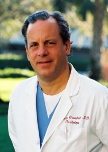
DrChaunceyCrandallDr. Crandall Chaucey: Health report on Chelation, August 2014

“Whether its called homeopathy, natural therapy or complementary treatment, there’s no doubt that alternative medicine is booming. Today, Americans spend more than $34 billion on these techniques, and surveys show that 1 in 3 Americans have tried some type of alternative medicine…”

Click the link above for the full text pdf.

Craig Gustafson: An Interview with L. Terry Chappell, MD

ChappellL. Terry Chappell, MD: Considering the Past and Promise of Chelation Therapy

Interview by Craig Gustafson

ALTERNATIVE THERAPIES, MAY/JUNE 2014 VOL. 20, 3

L. Terry Chappell, MD, is in private practice with Celebration of Health Association in the cities of Bluffton and Toledo, Ohio. He is board-certified in family practice, geriatrics, chelation therapy, pain management, and advanced longevity medicine. As past president of the International College of Integrative Medicine, past president of the American College for Advancement in Medicine, and as a volunteer assistant clinical professor of medicine at Wright State College of Medicine, he has taught chelation therapy and other integrative medicine to students and doctors from around the world. He has published widely on chelation therapy in scientific journals and in popu- lar books. He has served as a consultant for the National Institutes of Health on several occasions. He has devoted his practice to safe, natural techniques to improve function and help the body heal. He has assembled a high-quality nursing staff and recruited naturopath Bob Angus to work as a team to achieve the best results possible for each individual patient. (Altern Ther Health Med. 2014;20(3):56-60.)

Alternative Therapies in Health and Medicine (ATHM): Did you always know you wanted to be doctor? Was there a specific event in your life that pushed you in that direction?

Dr Chappell: No. When I went to college, I thought that would be about the last thing I was going to be. I had an upper-class roommate who convinced me to try a few class- es, and I tried it, liked it, and I found out I could stand the sight of blood so I went ahead.

ATHM: Where did you go to school, and how did your edu- cation affect your perspective on medicine?

Dr Chappell: I went to DePauw University in Greencastle, Indiana, for undergraduate school. It offered a liberal arts education; you get a good education with a wide back- ground. I think that is very helpful when you are a doctor. Then, I went to medical school at the University of Michigan. That is where my parents did some work, and later my

daughter went there. We have got a lot of connections with the University of Michigan.

ATHM: Were there any particular experiences that—or mentors who—influenced your development as a physician during your medical school years?

Dr Chappell: I would say it was more the broad experience. There was a movement in our medical school class to be of service to underprivileged people and I got involved with that. We established a free clinic, and that was a pretty important influence. I also met my wife through that; she was a nurse in that program. Student Health Organization was the name of it.

ATHM: How did you first come across complementary inte- grative medicine?

Dr Chappell: I first came across it through my wife, who was a registered nurse doing graduate work in psychiatric nurs- ing. Her training involved Lamaze natural childbirth, doing things as naturally as possible, and some nutritional support. That was my first real contact with it. I saw what you could do to relieve pain and stress by nondrug methods. I decided to learn about that. After that, my wife and I did some ser- vice in Appalachia. We found that many of the people there were suffering from pain and depression. They were pre- scribed pain pills and antidepressants, and were not getting anywhere. I looked for some alternatives to help them and they responded very well. They were very interested in it and got me excited about the whole process.

ATHM: What types of things did you find to help the people in Appalachia?

Dr Chappell: I started out by learning ear acupuncture as it is practiced in France. I also learned hypnosis and some basic nutritional tests and treatments. It worked out quite well.

L. Terry Chappell, MD: Considering the Past and Promise of Chelation Therapy Interview by Craig Gustafson

 CONVERSATIONS

This article is protected by copyright. To share or copy this article, please visit copyright.com. Use ISSN#1078-6791. To subscribe, visit alternative-therapies.com

Conversations With L. Terry Chappell, MD ALTERNATIVE THERAPIES, MAY/JUNE 2014 VOL. 20, 3     57

ATHM: How much nutritional background did you get in medical school?

Dr Chappell: About half an hour. I have always joked that I was fortunate to get only half an hour of nutrition training because I found out later that most of what they taught was wrong—so I did not have as much to forget.

ATHM: How did you incorporate nutritional and preventive medicine into your practice?

Dr Chappell: I first got into nutritional work actually as a secondary concern. It started when one of my patients kept after me and kept after me to learn more about chelation therapy. We were living in Ohio at that time, where my wife is from. He had people who were working for him who were traveling all the way from Ohio to West Virginia to get chelation treatments. He was seeing great results and he really wanted me to get into it. I was pretty skeptical at that time, but I eventually did agree to visit a couple of doctors who were giving it. I was really impressed by the patients’ stories—how much better they were getting with the treatments. I went to the American Institute for Medical Preventives— later called American College for Advancement of Medicine, or ACAM— so I could learn more about this chelation thera- py. ACAM offered a tre- mendous opportunity to learn about nutritional therapy of all kinds. That is when I really got into it. I found it important to be sure about the quality and the availability of nutritional products. I started ordering them and providing them to my patients—who reaped the benefits. It was a really good move. I went to an intensive seminar by Jonathon Wright, MD, and Alan Gaby, MD. They give these seminars every year or two in which they go into great detail about the scientific paces of nutri- tional therapy. It was particularly fun for me to learn from Jonathan Wright because he was a classmate of mine at the University

of Michigan. We had not seen each other for quite a while, so it was really great to get back together with him and learn about the great work that he was doing.

ATHM: What role does chelation play within the spectrum of preventative medicine?

Dr Chappell: The beauty of chelation is that it removes toxic metals, and toxic metals can interfere with normal function- ing of the body and contribute to disease including immune diseases, circulation problems, and degenerative diseases of all kinds. We find that these are very important to look for. If we find toxic metals, we treat with chelation. Over and above that, chelation helps with circulation, and we believe that if you can improve circulation, you can help most diseases.

ATHM: Are those benefits attributed to the action of a particular chelation agent, or are different sub- stances used?

Dr Chappell: Chelation means to grab ahold of metals and take them out of the body. Some of those are normal metals that you have to replace, but there are chelating sub- stances that have a great affinity for toxic metals such as lead, mercury, cadmium, and arsenic. We use different chelating materials depending on what the toxicity is and what the purpose of treat- ment is. For vascular dis- ease, the best evidence is for EDTA. That was prov- en very nicely by the Trial to Assess Chelation Therapy, or TACT, which was finished a couple of years ago.

ATHM: Tell us about TACT.

Dr Chappell: TACT began as a mandate from US Representative Dan Burton of Indianapolis. He held con- gressional hearings about why the National Institutes of Health, or NIH, was not funding research on chelation therapy even though so many smaller studies pointed to its effectiveness. Eventually the NIH agreed to do the research

This article is protected by copyright. To share or copy this article, please visit copyright.com. Use ISSN#1078-6791. To subscribe, visit alternative-therapies.com

Conversations With L. Terry Chappell, MD58     ALTERNATIVE THERAPIES, MAY/JUNE 2014 VOL. 20, 3

and they called for proposals. TACT was a major double-blind RCT of patients who either got chelation or placebo. They also looked at high-dose vitamins versus low-dose vitamins, and patients received these treatments in addition to their treat- ment for vascular disease. Everybody in the study had suffered a heart attack, so everone had known vascular disease. They looked for cardiac events over 3 to 5 years. The cardiac events included death, new heart attacks, strokes, and hospitalization for severe heart problems. The study wound up with 1708 patients from the United States and Canada. It was a very impressive study and it came out with positive results. This is exactly the type of study that is required of medications to get approval by the FDA and to get accepted in medical circles. With chelation, as it has been a controversial therapy for quite a while, there is still some reluctance to accept it—even though there is proof.

ATHM: Did you participate in TACT?

Dr Chappell: Yes. I was one of the people who originally testified in the congressional hearing, and I was also involved with evaluating the different research proposals that came in. Eventually I did become an investigator in the study, and I did treat patients. We had 39 patients in the study. It was a very good experience.

ATHM: Who was the lead author for that?

Dr Chappell: Tony Lamas, MD. Dr Lamas is a research car- diologist on the faculty of Columbia University and associ- ated with the University of Miami in Florida. He has done several major research projects in the field of cardiology. He is very well respected and he made sure that the study was done without significant problems. It was very interesting because it was one of the first studies that NIH has funded which had university medical centers participating in it along with experienced clinicians out in the field. That coop- eration was very positive.

ATHM: You mentioned that chelation has had controversy surrounding its use. Can you describe why?

Dr Chappell: Chelation was first reported for vascular dis- ease in Detroit by Dr Norman Clarke. Initially, there was a fair amount of interest in it from cardiologists in the United States; this was back in the late 1950s. There was one group of cardiologists that did a study on it, and their conclusion— even though their data was positive for chelation—was that it was not any better than existing treatment. Unfortunately, that eliminated the interest in chelation, and conventional research was no longer done for many years after that. In fact, a small group of alternative doctors continued to use the therapy because they saw really good benefits from it. Quite a number of minor studies were done that supported its use. The AMA and some medical groups became quite criti- cal of the therapy. Finally, the AMA demanded a major study

be done in the early 1980s. Of course, the doctors who were doing chelation did not have the resources to do a large double-blind study. They were treating patients who came to them for help and for treatment, not for a study. It was very difficult to get that study going, though there were several attempts to do it. One started at Walter Reed Hospital, a US Army hospital, but that one fizzled during the Iraq War because the physicians who were doing the study were called off to the war and were not able to complete it. There were other universities that tried to do the studies; they just could not get it done for various reasons. During all this time, medical boards and other medical associations issued statements against the therapy. It was controversial, no doubt, but the NIH finally funded the study.

ATHM: If early trials had shown that it was no more effective than the current treatments, wouldn’t there still be some advantage to using it—considering potential side effects of drug therapy—if it were equally effective?

Dr Chappell: Yes. That is a good point and certainly could be the case. However, I will emphasize that in TACT, in order to prove its effectiveness, all the patients in the study got conventional treatment, so the improvement that they showed in TACT was over and above the conventional treat- ment. It was statistically significant that you could decrease cardiac events pretty substantially with chelation therapy during that trial.

ATHM: Can you comment on the findings coming out of TACT that had to do specifically with diabetic cofactors or comorbidity?

Dr Chappell: That was probably the biggest surprise to con- ventional physicians. Back in the 1960s, there was some work that indicated that diabetics did the best of all the patients, but that was just a couple of isolated studies. In TACT, you could separate out the patients who did have diabetes. The improvement of the patients who had both chelation plus high-dose vitamins decreased the subsequent cardiac events for these patients by 51%. That is a huge num- ber over just a 3- to 5-year period—much more than the usual medications and other treatments that are given for diabetes. The researches who are looking at diabetes are very interested in this, and they are looking at possible further research that might examine this in more detail.

ATHM: Efforts have been made to limit heavy metals in homes and workplaces. If that is the case, why should chela- tion be so important in medicine currently?

Dr Chappell: There is a reduction in lead and mercury in the environment, but there is still quite a bit there. In fact, those metals are both in the top 4 or 5 pollutants in the United States. The chemical toxicities occur and they have been linked to many diseases. Even small amounts of lead and

This article is protected by copyright. To share or copy this article, please visit copyright.com. Use ISSN#1078-6791. To subscribe, visit alternative-therapies.com

Conversations With L. Terry Chappell, MD ALTERNATIVE THERAPIES, MAY/JUNE 2014 VOL. 20, 3     59

mercury can be significant in diseases. This has been shown in many research projects published in the last few years. Unfortunately, up until this time, there has not been a lot of research on treating patients with toxic metals. Small amounts of lead are still big factors in the produc- tion of vascular disease. Why there is such reluctance to treat these patients, I have no idea. It sure makes sense that if you have a toxic substance and you can remove it, then it could make a big difference to that patient.

ATHM: Here in Minnesota, when you think of mercury, you think of fish. But the potential for exposure to mercury is quite a bit greater than most people assume. Where are those environmental exposures to mercury coming from?

Dr Chappell: The biggest one—and still controversial—is from amalgam fillings in teeth. It has been documented that if you have amalgam fillings in your mouth, the metals in them are silver and mercury. If you chew, you can vaporize enough of the mercury so that it can be measured—and it can be high, even toxic levels—just after chewing gum. The mercury amalgam issue is a big one. In some countries those types of fillings are outlawed. Other countries have limited them, but there is still that exposure, because there are an awful lot of people who have amalgam fillings in their mouth. There are other places in the environment: One of the biggest is from burning coal for power. If you breathe polluted air containing mercury, then you are going to absorb some of that into your system. Fish is a big factor, too. If you eat a lot of fish, you can develop a toxic level of mercury.

ATHM: The federal government is pushing one other source into our homes through compact fluorescent bulbs and end- ing the manufacture of incandescent light bulbs.

Dr Chappell: Yes, that is a scary one. If you break a light bulb, that can be a dangerous exposure. You really should call a company that deals with toxic exposures to clean it up. If you try to clean that up yourself, you are going to get a big exposure to mercury.

ATHM: There are several other heavy metals that we need to be aware of in our environment, correct?

Dr Chappell: Right. Arsenic is a big one, and third-world countries are particularly high in arsenic. India, especially, has a huge arsenic problem. Aluminum. Others we are just beginning to understand; one of them is gadolinium. Gadolinium is in the dye that they use for MRIs. When we do a challenge test and look for toxic metals, we can tell whether that individual has ever had an MRI in their lifetime because of high gadolinium levels. Gadolinium is a toxic metal. Tin is another very common one that we see, and nickel, too. Cadmium is very toxic, often related to cigarette smoke, but is also airborne in the environment. We find fre-

quently that people with eye problems have lead or cadmium at high levels. Antimony is another one that can be very toxic. That has been shown in cardiomyopathies; they might have very high levels of antimony. We are just in the beginning stages of understanding the effects of all these toxic metals. Each indi- vidual metal is toxic, but 2 or 3 of them together multiplies  the effect and that has not been studied. We certainly think it is a good thing to get rid of them as much as we can. Memory problems: Of course, these have been going up astronomically. Lead, aluminum, and mercury can all con- tribute to memory problems and other neurologic diseases. Often times, if we have patients who have these diseas- es—memory problems, neurologic problems of various kinds, sometimes even cancers—we do a challenge test to see if toxic metals are present. We try to improve their immune system and their neurologic function by removing the metals.

ATHM: You have written about testing for toxic metals and that some of the more obvious tests that have been used in the past may not be tremendously effective or may even be  misleading. How so?

Dr Chappell: The gold standard for toxic metals is to do a blood test. The problem is that these toxic metals do not stay in the blood very long. If you are working around them every day and worried you are getting a toxic load, this is fine because you will find elevated levels if there has been a recent exposure. In the body, these metals are stored in the bone, in the brain, and in the fat cells. They are not floating around in the blood. The best way to look for toxic metals is to do a challenge test, where you give a chelating substance and then you mea- sure the urine, or sometimes the stool, to see how many toxic metals you pull out over the next few hours. That is the best way to see if there is a toxic load. This can be really impor- tant. For example, if you have lead in the bone, and you have a bone injury or even maybe a bone surgery, like with a joint replacement, you are going to have leakage of the contents of that bone into the rest of the body and you might get a flood of lead that has been stored in that bone. That could cause adverse effects after a fracture or a surgery that could be very significant.

ATHM: Individuals are engaging in some pretty aggressive detoxification procedures, often without practitioners over- seeing their program. Could this lead to a massive release of heavy metal toxins within the body?

Dr Chappell: Possibly, yes. They have not been well studied, to my knowledge, but I would be concerned about that. You also have to be careful that whoever is doing it is knowledge- able about the chemistry of the substances. For example, EDTA is a very safe substance that is found in tiny amounts in cereals in our grocery store. In higher doses it is used as a chelating substance to pull out toxic metals. Fortunately, it

This article is protected by copyright. To share or copy this article, please visit copyright.com. Use ISSN#1078-6791. To subscribe, visit alternative-therapies.com

Conversations With L. Terry Chappell, MD60     ALTERNATIVE THERAPIES, MAY/JUNE 2014 VOL. 20, 3

becomes tightly bound to toxic metals and is excreted quick- ly. Only about 5% of the EDTA is absorbed orally. You are not going to get a very predictable detoxification with the EDTA given by mouth. Another problem with poor absorbtion of oral EDTA is that it might bind with normal metals and prevent their absorption. So you could get malnourished even though you are eating foods containing the normal metals that you need. It can be controversial. It can be difficult. It has potential for doing harm to a patient, if the person who is doing the treat- ment is not knowledgeable.

ATHM: How about a program where the contents of fat cells are flushed rapidly? They’re losing a tremendous amount of weight in a short amount of time and releasing the contents of those fat cells into the bloodstream.

Dr Chappell: I think that would be an important consider- ation, as well. You could possibly get a flood of toxic metals by releasing them from the contents of fat cells.

ATHM: Considering unavoidable environmental exposures, should there be some level of heavy metal detoxification built into your lifestyle?

Dr Chappell: Certainly regular exercise, particularly if you can build up to a sweat; that is a very good way to detoxify, gradu- ally. Another route is through your digestive tract. If you eat high-fiber foods, you might also benefit from detoxification and prevention of absorbing toxicants from your food supply. Generally with chemicals—and there have been many chemi- cals that have been identified that can be toxic to the body—the body has mechanisms to detoxify and handle those chemicals, but sometimes it does not keep up. I find that milk thistle is a good herb that can be used to enhance the detoxification effect.

ATHM: What about trying to support your body’s antioxi- dant system, like precursors for glutathione?

Dr Chappell: Glutathione is a wonderful antioxidant the body needs. It is essential for many bodily functions. N-acetylcysteine, or NAC, is one precursor that can be used, as well as vitamin E and selenium to help with the produc- tion of glutathione. Glutathione by itself is not very well absorbed into the body, so it is hard to get high levels that way, although there has been some research recently saying that if you use a liposomal preparation you can get much better absorption. There are more products being developed all the time to enhance antioxidant effects, and they are important defense mechanisms.

ATHM: You are currently trying to get the word out about a grant program for chelation therapy. Please describe the program and provide the details for applying.

Dr Chappell: In order to get complete acceptance of chela- tion for toxic metals as well as the use of EDTA for vascular disease, there needs to be another study to support TACT, which was just completed and published—although certain parts of it are still being published. There are not many orga- nizations right now that sponsor research in this area. The NIH is the major one that is a potential sponsor. However, in order to get a major contributor to finance a big study, a lot of work needs to be done. That involves preliminary work, study planning, statistical projections, and so forth. The International College of Integrated Medicine, or ICIM, which teaches doctors how to do chelation safely and effectively, decided to put forward money to stimulate that process. The ICIM has sent out a call for proposals for any- body who is interested in doing chelation therapy. ICIM will give a grant of $20 000 for putting together a proposal for a larger grant and study that can replicate the findings of TACT. This announcement has been received with a lot of enthusiasm, and we expect to get innovative proposals of different diseases that we can test that may support TACT. For example, one thing that has been proposed concerns diabetic patients who have been told that they need an amputation. A study could see if EDTA can prevent amputa- tions. There was a smaller study that actually did show that effect in Denmark. The deadline for the grant is May 31, and ICIM would like to receive as many proposals as possible.

ATHM: Where should the proposals be sent, and how?

Dr Chappell: The proposal should be sent to  wendy@icimed.com. Any questions could be directed to her as well. She is the executive director of ICIM.

Intravenous Vitamin C Saves NZ Man with Flu Damaged Lung

chest-xray-1526779-639x626A 56 year old male was referred to Auckland Hospital ICU on 1 July 2009 with total respiratory failure, for ECMO external oxygenation. The patient had contracted H1N1 Swine flu (confirmed by tests) while on holiday overseas, and had developed what is known as ‘white out’ pneumonia. This refers to x-rays showing no air space in the lungs.

After 20 days of life-sustaining ECMO treatment and other critical care, the patient, who was unconscious by induced coma, had not responded. The ICU team advised the family of the likely outcome and had prepared them for the possibility of the patient’s death.

Family members approached Centre for Advanced Medicine Limited (CAM) for advice on the clinical use of intravenous vitamin C for such cases.

At the family’s request, information was provided to ICU doctors including ISO 9001:2008 registered protocols, safety data, dosages and access to vials of IV vitamin C under CAM’s license for wholesale medicines.

The ICU team agreed to administer intravenous vitamin C according to the family’s wishes. This decision acknowledged the family’s rights, in compliance with the New Zealand Health and Disability Act, 1997.

The patient received intravenous vitamin C starting on the evening of 21 July, continuing until 29 July. 25 grams was provided on the first day increasing over the first three days to 50 grams twice daily which was sustained for a further six days.

By 24 July x-rays indicated increasing lung function and ECMO external oxygenation was discontinued on 26 July. After several days of assisted ventilation and critical care for ongoing secondary conditions, the patient was able to commence his recovery and rehabilitation. The patient was discharged from hospital on Friday 18 September, and is recovering at home on the farm.

The decision by the Auckland Hospital ICU team to administer adequate dosages of IV vitamin C, and their skillful coordination of ICU procedures, were responsible for the positive medical outcome.

Permission from the patient and his family has been sought by CAM to publish these details on its website and elsewhere in the interests of accuracy. This permission was willingly provided and  CAM expresses its thanks, admiration and respect. CAM welcomes opportunities to provide similar professional support for registered medical practitioners and their patients.

Obtained from:

http://centerhealingarts.com/treatment-programs/iv-therapy-nutrition-chelation/intravenous-vitamin-c-saves-nz-man-flu-damaged-lung/

Protocol Controversies for Treating Cardiovascular Disease with EDTA Chelation Therapy

L. Terry Chappell and Jeanne A. Drisko, Reprinted from the Townsend Letter with permission

IntroductionjDrisko

The Trial To Assess Chelation Therapy (TACT) is the only large, randomized clinical trial to provide statistically significant evidence that EDTA chelation therapy with high-dose multivitamins can reduce future cardiac events in patients with known cardiovascular disease(1,2).  TACT utilized the published protocol(3) that is used by organizations such as the American College for Advancement in Medicine (ACAM), the International College of Integrative Medicine (ICIM), the American Board of Clinical Metal Toxicology (ABCT), and the International Board of Clinical Metal Toxicology (IBCMT), all of which teach physicians how to administer the therapy and/or test them to provide certification.

TACT used an intravenous dose of 3 Gm of disodium EDTA with magnesium, adjusted downward if kidney function was compromised, 7 Gm of vitamin C, 500 cc of sterile water, and several minor additives, all infused over a minimum of 3 hours(1,2). See Table 1. The published protocol is more flexible, allowing for 1.5 Gm-3 Gm of disodium magnesium EDTA over no more than 1 Gm per hour and varying amounts of vitamin C, as long as the osmolality of the treatment solution is not hypotonic and not so hypertonic as to cause problems.  Calcium EDTA has also been used in various forms with claims of effectiveness for vascular disease.  The use of Calcium EDTA, especially in the oral form, to treat cardiovascular disease has been criticized by the teaching organizations mentioned above.  Concerns have also been raised about high doses of vitamin C, which becomes a pro-oxidant instead of an anti-oxidant at certain levels(4).

The purpose of this article is to discuss the rationale, evidence, and experience of physicians who are acknowledged experts in the use of EDTA for treating cardiovascular disease.  We hope to clarify whether Calcium EDTA should be used to treat vascular disease and how much EDTA and vitamin C are effective and safe to use.

The Published Protocol

 TACT used the 3 Gm basic dose of disodium EDTA with magnesium to treat patients who had a history of documented myocardial infarction.  The basic protocol for TACT is shown in Table 1.  {Insert Table 1} The 3 Gm dose for disodium EDTA has been taught for years, and many doctors who provide intravenous chelation therapy use it routinely.  However, there is evidence that a lesser dose might be just as effective(6,7,8,9).  As a result, a substantial number of treating physicians use the lesser dose, based on these reports.  Obviously, a lesser treatment time is more convenient for patients.  Neither dose puts the kidneys at risk as long as the required rate of administration is followed.  For patients with congestive heart failure, a lesser fluid volume for the treatment might be advantageous.

Chappell and Stahl performed a meta-analysis of studies showing objective improvement for patients with cardiovascular disease treated with intravenous EDTA chelation therapy(5).  Nineteen published studies involving 22,765 patients met the inclusion criteria.  All of these studies used the 3 Gm dose of EDTA with one exception.  Olszewer and Carter treated 2,482 patients with the 1.5 Gm dose, and 2,379 improved(6).  In the meta-analysis, 87% of patients improved, and there was a correlation coefficient of 0.88 between improvement in vascular function and treatment with EDTA.  Patients of the physician who used the 1.5 Gm dose did as well as those from the other sites combined.

Chappell and associates did a follow-up meta-analysis of 32 unpublished reports on 1241 patients(7).  1086 or 88% showed measurable improvement. 778 patients were treated with the 1.5 Gm bottle.  A comparison of the 1.5 Gm and 3 Gm doses in this study showed almost identical results. 

Born and Geurkink published a retrospective, randomized study comparing patients with peripheral artery disease treated with the 3 Gm dose of EDTA to those treated with the 1.5 Gm dose(8).  20 treatments were given to 15 patients in each group.  Those treated with the lower dose improved using Doppler ultrasound by an average of 123%.  The patients in the 3 Gm group improved by an average of 70%.  The results were statistically significant.  One patient treated with 1.5 Gm improved 715%.  That patient was omitted from the study as an outlier.

Chappell and associates compared 220 vascular patients treated with a basic course and maintenance chelation to matched controls from the literature(9).  An average of 58 treatments were given. Subsequent cardiac events were much less in the EDTA-treated group.  The patients treated with the 1.5 Gm dose had virtually the same results as those with the 3 Gm dose.

An in vitro study published in Surgery in 1962 showed the mobilization of calcium from atherosclerotic plaque with EDTA in the laboratory(10).  The results demonstrated that the longer the tissue is exposed to EDTA, the more calcium was removed.  To our knowledge, this finding has not been confirmed in vivo.

Blaurock-Busch observes that the German Chelation Society approves both a 2 Gm and 3 Gm dose(11).  Gordon wrote the first American Academy of Medical Preventics (AAMP) chelation protocol in 1972, based on the work of such pioneers as Clark and Lamar.  It was not published, but it was used in coursework for many years.  He listed the EDTA dose of 50 mg/ Kg.  Cranton’s 1989 Textbook refers to a maximum of 3 Gm dose, except for large patients who could receive up to 5 Gm at 50 mg/Kg.  The textbook was updated in 2001(12).  Rozema’s protocol for EDTA(3) lists both a 3 Gm and 1.5 Gm dose, as does van der Schaar’s 2012 Textbook(13).  The latter has a maximum of 4 Gm for large patients.  All of these protocols insist on an infusion rate of disodium EDTA not faster than 1 Gm per hour to avoid overloading the kidneys.

Because of TACT, the best evidence for treatment of vascular disease with intravenous disodium EDTA lies with the 3 Gm dose.  However, the published studies cited above that compare the 3 Gm dose to the 1.5 Gm dose show the latter to be as effective.  As noted, one study showed the 1.5 Gm dose to be more effective for peripheral vascular disease.  Future large clinical trials will be necessary to determine the lowest amount of EDTA that can produce the best outcome in cardiovascular disease.

Mechanisms of action for EDTA

Proposed mechanisms of action for EDTA chelation therapy have been documented(14), but no consensus exists as to which mechanism(s) are most important to treat vascular disease.  It is well known that both disodium EDTA and calcium EDTA can remove heavy metals.  Such metals as lead, cadmium, and mercury increase the risk of vascular diseases by increasing free radical activity(15).  Reduction of free radicals by EDTA infusions reduces inflammation, which might lesson the likelihood of the rupture of unstable plaques(16).  The clot that occurs as a result of this rupture is the accepted mechanism for most myocardial infarctions and strokes.  A small study by Chappell and Angus showed a reduction of brachial artery stiffness with chelation(17).  Iron deposits have been found in macrophage foam cells, which further increase free radicals and inflammation.  Excessive copper also increases free-radical activity.  EDTA chelates both iron and copper(18).

Lowering blood calcium levels with intravenous boluses of disodium EDTA can inhibit platelet aggregation for weeks at a time(19).  Intravenous EDTA has been proposed as a safer substitute for clopidogrel to prevent clotting after inserting drug-eluting stents(20).  The anti-clotting effect is likely to be an important mechanism for chelation’s cardiovascular benefits.  Selye demonstrated harmful deposition of calcium in soft tissue when a sensitized individual is exposed to a new challenge after a suitable interval(21). The drop in serum calcium that occurs almost immediately upon IV infusion of disodium EDTA stimulates parathyroid activity.  Parathormone mobilizes calcium from soft tissue deposits, but the effect is irregular.  Although there are case reports that plaque can be reduced with disodium chelation(22,23), studies have not shown a predictable improvement in lumen size for arteries blocked with plaque.  It is possible that the calcium reduction cascade stabilizes vulnerable plaques, but this also has not been proven. 

High doses of magnesium are put into the intravenous treatment solution, which prevents adverse effects from the brief drop in calcium levels.  Improved levels of intracellular magnesium might reduce irritable foci that cause arrhythmias and lower blood pressure. To prevent progressive calcium depletion, it is important that IV infusions of disodium EDTA be given no more often than 2-3 days per week, with at least 24 hours between treatments.  With 60 years of use of intravenous disodium EDTA for vascular disease, no fatalities have been attributed to EDTA when the protocol has been followed.  However, there have been isolated fatalities when disodium EDTA was administered by rapid IV push.

Nitric oxide(NO) is an important signaling molecule that is anti-atherosclerotic.  NO production declines with age and is worse with a high fat diet.  Lead inhibits NO formation.  EDTA not only removes lead but also independently increases NO production(24).  This might be an important mechanism for improved circulation for both disodium EDTA and calcium EDTA.

Vitamin K2 also might help remove metastatic calcium from arterial walls.  It has been suggested as an oral supplement to augment the decalcifying effect of disodium EDTA(25).  However, vitamin K2 is not currently included in the chelation therapy protocol

Calcium EDTA

Intravenous calcium EDTA is approved for removing lead and is used to treat accumulations of other toxic metals.  Since there is no reduction of serum calcium as is seen with disodium EDTA, certain mechanisms that are proposed for treating vascular problems do not apply.  Specifically, metastatic calcium is not mobilized and platelets are not inhibited.

 Oral, sublingual, transdermal, and rectal EDTA all consist of calcium EDTA. Oral EDTA is only about 5% absorbed.  Rectal EDTA might be absorbed as much as 35-37% (26).

Intravenous calcium EDTA is used widely as a challenge test and a treatment for toxic metals.  It was used in a small study by Lin that showed that non-diabetic patients with moderate kidney disease might progress less rapidly with EDTA treatment than without(27).  Chen and associates showed that diabetic nephropathy in the presence of high lead levels progressed at a slower rate than controls when their lead levels were reduced and kept under control with 1 Gm calcium EDTA treatments IV(28).  High levels of lead have been shown to be associated with lower blood pressure and an increased risk of vascular disease(29).  Reducing the lead burden might result in improved blood pressure and better circulation to the kidneys.  However, without a drop in serum calcium, decalcification of the arterial wall is highly unlikely.  The many published studies showing improvement in vascular disease, including TACT, all have used disodium EDTA with magnesium.

Garry Gordon has proposed that calcium EDTA combined with lecithin and other nutrients improves blood viscosity, and he cites the work of Lowe (30) and others.  One might expect this to be the case since lavender-top tubes with EDTA are used to anti-coagulate blood drawn from patients for testing.  However, the EDTA used for that purpose is potassium EDTA (K2EDTA), not calcium EDTA.  We were unable to find evidence that calcium EDTA reduces platelet activity directly.  One mechanism for inhibition of platelet aggregation is a depletion of calcium ions.  However, another probable mechanism that applies to calcium EDTA is its stimulation of the production of NO.  Several oral nutrients that can lesson platelet aggregation, such as vitamin E and gingko, can be given orally along with calcium EDTA.

Cranton points out a potential danger of oral chelation on his website(31).  Some toxic metals that are ingested might not be absorbed into the body if calcium EDTA is present, but many more essential minerals will also not be absorbed.  Depletion of zinc, chromium, copper, manganese, and other minerals can reduce antioxidant defenses and endocrine function.  Cranton stresses the importance of the rapid decrease in both toxic metals and calcium with disodium EDTA.  This occurs extracellularly since EDTA does not enter the cells.  A re-equilibration results so that calcium is mobilized as described above and toxic metals are brought out of storage in the bone, brain, and fat cells.

Calcium EDTA is widely sold and advertised as an ingredient in various nutritional supplements.  Claims of effectiveness for calcium EDTA in treating vascular disease are often made based on research that was done for intravenous disodium EDTA.  Calcium EDTA and disodium EDTA are two separate compounds that act on calcium differently in the body.  Although useful mechanisms of action might apply for calcium EDTA, we did not find any clinical trials that support the use of calcium EDTA for treating vascular problems.

Van der Schaar’s textbook describes many toxic metals and chelating agents(13).  DMSA, DMPS, and the two forms of EDTA are commonly used in clinical practice at this time.  DMSA is available orally and is used to chelate lead and mercury in adults and children.  DMPS is a compounded substance for oral or IV use, mostly for lead and mercury, but it is not an FDA-approved medication.  DFO is sometimes used for iron overload parentally but serial phlebotomies are generally more effective.  D-penicillamine can be helpful as a challenge test and occasional treatment.  These medications can be used in combination if the prescribing physician is experienced. The two forms of EDTA are broader chelators and are especially effective for lead.  EDTA has perhaps the weakest affinity for mercury.  If mercury is elevated with a challenge test, it might be prudent to treat with DMSA or DMPS before prescribing intravenous EDTA.  Maintaining good levels of beneficial minerals is important no matter what chelation agent(s) is/are prescribed.  Treatment with DMSA or DMPS reduces free radical activity by binding and excreting heavy metals, which might be beneficial.  However, we did not find any clinical trials that have studied either one as a treatment or preventative for vascular disease.

Vitamin C

As with any medical practice, whether conventional or alternative, different approaches and individualized styles of practice evolve. Some of the differences in approaches relate to experience and some are based on growing evidence from the scientific literature. And so it is with EDTA chelation therapy with certain groups giving differing amounts of EDTA over varying times and by different routes of administration, while others advocate using different formulations and combinations of additives in the mix. One proposed change has been the recent suggestion that vitamin C or ascorbic acid be removed from EDTA chelation therapy(4,32) because of its known action as a pro-oxidant in the extracellular space in living systems(33,34).

Seminal findings regarding the unexpected pro-oxidant action of intravenous vitamin C were discovered in the National Institutes of Health (NIH) lab of Mark Levine, MD along with his colleagues, Qi Chen, PhD and others(33,34).  Levine and colleagues clearly defined that oral vitamin C was a vitamin with tight physiologic control and anti-oxidant properties, while intravenous vitamin C administration bypassed tight control and through Fenton chemistry became pro-oxidative in the extracellular space(35-37). In people with normal G6PD status, the pro-oxidative nature of vitamin C does not occur in the vascular space(34).

It is interesting that another well-known anti-oxidant, glutathione, does not behave like vitamin C when injected in high doses(38).  Glutathione maintains its anti-oxidant properties even when injected at increasing concentrations and has led to the recommendation against adding IV vitamin C and IV glutathione together at the same setting(38).  To date, other anti-oxidants such as alpha lipoic acid have not been evaluated in this manner to determine if they might exhibit a dual nature like vitamin C.

The pro-oxidative nature of intravenous vitamin C has led some to postulate that adding vitamin C to EDTA chelation therapy might have a deleterious effect on patients with already high oxidative burden, as seen in diabetes(4,32).  The hypothesis is that patients with oxidative disease processes may not be able to tolerate the additional oxidative burst that briefly occurs after intravenous vitamin C.  Roussel and colleagues conducted a small uncontrolled trial in 6 adults where EDTA chelation therapy was administered according to standard protocol except for elimination of the vitamin C from the infusate(4).  In the reported trial, markers for oxidative damage were evaluated in the absence of added vitamin C and were found not to be present. The group concluded that EDTA chelation therapy without added vitamin C decreases oxidative stress. But as Roussel and colleagues clearly stated, the small trial was not designed to test the curative effects of the chelation therapy. They acknowledged they were focusing solely on anti-oxidative effects.

The Roussel trial is in contrast to the TACT trial where 1,708 participants with known cardiovascular disease were enrolled(2).  The sample size was chosen so that the effect of EDTA chelation therapy on cardiovascular outcomes could be evaluated. The standard accepted protocol was chosen and this included 7 grams of vitamin C injected at each infusion(1).  An unexpected and remarkable finding at the conclusion of the TACT trial was the marked reduction in cardiovascular events in diabetic participants (2).  This has prompted the NIH to ask researchers to focus on the positive effects in the standard EDTA infusate that may have promoted such beneficial outcomes(39). However, the belief that intravenous vitamin C is a harmful pro-oxidant has led other experienced practitioners of chelation therapy to abandon the addition of vitamin C to the mix(32).  The trial findings with the small sample size and the concerns raised by Roussel and colleagues are contradicted by the positive outcomes of TACT.

What then is the effect that vitamin C plays in the chelation infusate? Is it related to the pro-oxidative burst? Is it related to other as of yet undescribed effects, apart from Fenton chemistry? It is advisable to remember that in the vascular space, when there is normal G6PD status, there are no significant detectable levels of hydrogen peroxide and no detectable pro-oxidant effect(33,34,40).  Any hydrogen peroxide that might be formed after infusion of vitamin C is quickly and effectively quenched in the vascular space, unlike what occurs in the extracellular space. Is it possible that vitamin C at increased concentrations has an effect on the endothelium? Or on the function of blood elements like the red blood cells that are so critical for oxygen mobilization? Unpublished research carried out by the Levine team points to this possibility.

The epidemiology literature shows that vitamin C is critical for improvement of HbA1c and avoiding untoward effects in diabetics(41).  However, it can easily be argued that this is a vitamin effect not a pharmacologic effect. In the tobacco literature it has been shown that the prolonged and destructive exposure to tobacco smoke markedly reduces available vitamin C levels in vivo that are only replenished effectively with intravenous infusion(42-46).  Functional effects on the microvascular bed can be reversed with IV vitamin C(43,45).  It can be argued that tobacco smoking is a model for highly oxidative chronic diseases such as diabetes.  Benefits of infused vitamin C could have positive effects on microvasculature function. This would certainly be a good model for future research.

Other nonvascular effects of IV vitamin C also come into play such as the effects of ascorbate in steroidogenesis, vascular tone, adrenal gland function during stress, and general well-being(47-50).  Taking the narrow view that vitamin C acts only a pro-oxidant or an anti-oxidant may result in the risk of excluding vitamin C with its various positive functions, both known and as yet unknown, in the beneficial treatment of cardiovascular disease with EDTA chelation therapy.

Conclusions

Scientific evidence, especially with TACT, supports intravenous disodium EDTA with magnesium, along with oral multivitamins, to treat vascular disease.  Disodium EDTA can only be given by slow intravenous drip, at a rate no faster than 1 Gm per hour.  Under no circumstances can this preparation be given by intravenous push because of its effect to rapidly lower serum calcium.  Evidence supports treatment doses of 1.5 or 3.0 Gms of disodium EDTA.  The dose should be reduced if kidney function is impaired.  Kidney function should be monitored during the course of treatment. Treatments should be limited to no more than 2 or 3 days per week with at least 24 hours between treatments.  20-30 treatments are needed to complete a basic course of treatment for vascular disease(3).  More treatments may be required in difficult cases.  Most experts recommend monthly maintenance after the basic course is completed.  If the published protocol is followed, safety is not an issue(2).  Disodium EDTA with magnesium effectively removes heavy metals from the body.  Other likely mechanisms of action include reduced platelet aggregation, mobilization of metastatic calcium by parathormone, increased NO production, and antioxidant activity.

Calcium EDTA can be given intravenously or by other routes of administration to remove toxic metals.  Oral absorption is only 5% and rectal absorption might be as high as 35-37%.  Calcium EDTA does not have all the mechanisms of action that disodium EDTA does to reduce or prevent vascular disease.  However, calcium EDTA with multivitamins increases NO production and decreases free radical activity.  Moderately impaired kidney function might improve with IV Calcium EDTA.  Clinical trials have not been done to support calcium EDTA as a treatment for vascular disease at this time.  Calcium EDTA should not be given on a continuing basis without being careful to avoid depletion of essential mineral nutrients. Optimal mineral balance might be difficult to accomplish with oral preparations given on a daily basis.

At this juncture because of the positive TACT outcomes, vitamin C should not be excluded from or reduced in the infusate. The hypothesis that IV vitamin C results in a significant deleterious oxidative burst has not been born out and as part of the total chelation component seems to provide an additional benefit in patients with diabetes and cardiovascular disease. As shown in other conditions with high oxidative environments, IV vitamin C can provide protection and vascular stability. Exciting research opportunities in parsing out the effect of the various chelation components in treating cardiovascular disease lie ahead. 

*See responses from physicians below References section

References    

  1. Lamas G, Goertz C, Boineau R, Mark DB, Rozema T, Nahin RL, Drisko J, Lee KL. Design of the Trial to Assess Chelation Therapy (TACT). Am Heart J [Internet]. Mosby, Inc.; 2012 Jan [cited 2013 Aug 12];163:7–12.
  2. Lamas GA, Goertz C, Boineau R, Mark DB, Rozema T, Nahin RL, Lindblad L, Lewis EF, Drisko J, Lee KL. Effect of disodium EDTA chelation regimen on cardiovascular events in patients with previous myocardial infarction: the TACT randomized trial.  JAMA 2013;309:1241-1250.
  1. Rozema TC. Special issue: protocols for chelation therapy.  J Adv Med 1997;10;5-100.
  2. Roussel AM, Hininger-Favier I, Waters RS, Osman M, Fernholz K, Anderson R. EDTA chelation therapy, without added vitamin C, decreases oxidative DNA damage and lipid peroxidation. Altern Med Rev [Internet]. 2009 Mar;14(1):56–61.
  3. Chappell LT, Stahl JP.  The correlation between EDTA chelation therapy and improvement in cardiovascular function: a meta-analysis.  J Adv Med 1993;6:139-63.
  4. Olszewer E, Carter JP.  EDTA chelation therapy in chronic degenerative disease.  Med Hypoth 1988;27:41-49.
  5. Chappell LT, Stahl JP, and Evens R.  EDTA chelation treatment for vascular disease: a meta-analysis using unpublished data.  J Adv Med 1994;7:131-142.
  6. Born GR, Geurkink TL.  Improved peripheral vascular function with low dose intravenous ethylene diamine tetraacetic acid (EDTA).  Townsend Letter 1994;July:722-726.
  7. Chappell LT, Shukla R, Yang J, Blaha R, et al.  Subsequent cardiac and stroke events in patients with known vascular disease treated with EDTA chelation therapy: a retrospective study.  Evid Based Integrative Med 2005;2:27-35.
  8. Wilder LW, et.al. Mobilization of atherosclerotic plaque calcium with EDTA utilizing the isolation-perfusion.  Surgery;52.
  9. Blaurock-Busch E.  Toxic metals and antidotes: the chelation therapy handbook.  Germany, MTM Publishing, 2010.
  10. Cranton EM, ed.  A textbook on EDTA chelation therapy, second edition.  Charlottesville, Va, Hampton Roads, 1989, updated 2001.
  11. Van der Schaar PJ, et.al.  Clinical metal toxicology, 10th edition.  International Board of Clinical Metal Toxicology. Leende, The Netherlands 2010.
  12. Olmstead SF.  A critical review of EDTA chelation therapy in the treatment of occlusive vascular disease.  Klamath Falls, Oregon, Merle West Medical Center Foundation, 1998.
  13. Halstead BW and Rozema TC.  The scientific basis of EDTA chelation therapy, second edition, Landrum, SC, TRC Publishing, 1997.
  14. Heistad DD.  Unstable coronary plaques.  N Engl J Med 2003;349:2285-2288.
  15. Chappell LT, Angus RC and associates.  Brachial artery stiffness testing as an outcome measurement for EDTA-treated patients with vascular disease.  Clinical Prac Alt Med 2000;1:225-229.
  16. Cranton EM, Frackelton JP.  Free radical pathology in age-associated diseases: treatment with EDTA chelation, nutrition, and anti-oxidants.  J Hol Med 1984;6;6-37.
  17. Zucker MB, Grant RA.  Nonreversible loss of platelet aggregability induced by calcium deprivation.  Blood. 1978;52:505-13.
  18. Chappell LT.  Should EDTA chelation therapy be used instead of long-term clopidogrel plus aspirin to treat patients at risk from drug-eluting stents?  Altern Med Rev. 2007;12:152-158.
  19. Selye, H.  Calciphylaxsis. University of Chicago Press, Chicago 1962.
  20. Rudolph CJ, McDonagh EW.  Effect of  EDTA chelation and supportive multivitamin/mineral supplementation on carotid circulation: case report.  J Adv Med 1990;3:5-12.
  21. Rubin M.  Magnesium EDTA chelation, chapter 175 in Cardiovascular Drug Therapy, editor Messerli FH.  WB Saunders Co, Philadelphia 1996.
  22. Barbosa F, Sertorio JTC, Garlach RF, Tanus-Santos JE.  Clinical evidence for lead-induced inhibition of nitric oxide formation. Arch Toxicol 2006;80:811-816.
  23. Rheaume-Bleue K.  Vitamin K2 and the calcium paradox.  John Wiley & Sons Canada, Ltd.  Mississauga, Ontario 2012.
  24. Ellithorpe, R.  Comparison of the absorption, brain, and prostate distribution, and elimination of CaNa2EDTA of rectal chelation suppositories to intravenous administration.  JANA 2007;10:38-44.
  25. Lin JL, Lin-Tan DT, Hsu KH, et al.  Environmental lead exposure and progression of renal diseases in patients without diabetes.  N Engl J Med. 2003;348:277-86.
  26. Chen Kuan-Hsing and associates.  Effect of chelation therapy on progressive diabetic nephropathy in patients with type 2 diabetes and high-normal body lead burdens.  Am J Kidney Dis 2012;60:530-538.
  27. Nash D, Magler L, Lustberg M, et.al.  Blood lead, blood pressure, and hypertension in perimenopausal and postmenopausal women.  JAMA 2003;289:1523-1532.
  28. Lowe GD and associates.  Blood viscosity and risk of cardiovascular events: the Edinburgh Artery Study.  Br J Haematol 1997;96:168-173.
  29. Cranton EM.  Http.//drcranton.com/chelation/oralchelation.htm. accessed Dec 30, 2013.
  30. Born T, Kontoghiorghe CN, Spyrou A, Kolnagou A, Kontoghiorghes GJ. EDTA chelation reappraisal following new clinical trials and regular use in millions of patients: review of preliminary findings and risk/benefit assessment. Toxicol Mech Methods [Internet]. 2013 Jan [cited 2013 Dec 29];23:11–7.
  31. Chen Q, Espey MG, Krishna MC, Mitchell JB, Corpe CP, Buettner GR, Shacter E, Levine M. Pharmacologic ascorbic acid concentrations selectively kill cancer cells: action as a pro-drug to deliver hydrogen peroxide to tissues. Proc Natl Acad Sci U S A [Internet]. 2005 Sep 20;102:13604–9.
  32. Chen Q, Espey MG, Sun AY, Lee J-H, Krishna MC, Shacter E, Choyke PL, Pooput C, Kirk KL, Buettner GR, Levine M. Ascorbate in pharmacologic concentrations selectively generates ascorbate radical and hydrogen peroxide in extracellular fluid in vivo. Proc Natl Acad Sci U S A [Internet]. 2007 May 22 [cited 2012 Oct 29];104:8749–54.
  33. Levine M, Conry-Cantilena C, Wang Y, Welch RW, Washko PW, Dhariwal KR, Park JB, Lazarev A, Graumlich JF, King J, Cantilena LR. Vitamin C pharmacokinetics in healthy volunteers: evidence for a recommended dietary allowance. Proc Natl Acad Sci U S A [Internet]. 1996 Apr 16 [cited 2013 Mar 22];93:3704–9.
  34. Padayatty SJ, Levine M. New insights into the physiology and pharmacology of vitamin C. CMAJ [Internet]. 2001 Feb 6;164:353–5.
  35. Padayatty SJ, Sun H, Wang Y, Riordan HD, Hewitt SM, Katz A, Wesley RA, Levine M. Vitamin C pharmacokinetics: implications for oral and intravenous use. Ann Intern Med [Internet]. 2004 Apr 6 [cited 2013 Mar 22];140:533–7.
  36. Chen P, Stone J, Sullivan G, Drisko J, Chen Q. Anti-cancer effect of pharmacologic ascorbate and its interaction with supplementary parenteral glutathione in preclinical cancer models. Free Radic Biol Med [Internet]. 2011 Aug 1 [cited 2013 Apr 12];51:681–7.
  37. http://nccam.nih.gov/news/2013/111913?nav=gsa, accessed 12/30/2013.
  38. Chen Q, Espey MG, Sun AY, Pooput C, Kirk KL, Krishna MC, Khosh DB, Drisko J, Levine M. Pharmacologic doses of ascorbate act as a prooxidant and decrease growth of aggressive tumor xenografts in mice. Proc Natl Acad Sci U S A [Internet]. 2008 Aug 12;105:11105–9.
  39. Kositsawat J, Freeman VL. Vitamin C and A1c relationship in the National Health and Nutrition Examination Survey (NHANES) 2003-2006. J Am Coll Nutr [Internet]. 2011 Dec;30:477–83.
  40. Alberg A. The influence of cigarette smoking on circulating concentrations of antioxidant micronutrients. Toxicology [Internet]. 2002 Nov 15;180:121–37.
  41. Ambrose J, Barua RS. The pathophysiology of cigarette smoking and cardiovascular disease: an update. J Am Coll Cardiol [Internet]. 2004 May 19 [cited 2013 Nov 13];43:1731–7.
  42. Northrop-Clewes C, Thurnham DI. Monitoring micronutrients in cigarette smokers. Clin Chem Acta [Internet]. 2007 Feb [cited 2013 Dec 9];377(1-2):14–38.
  43. Schindler TH, Nitzsche EU, Munzel T, Olschewski M, Brink I, Jeserich M, Mix M, Buser PT, Pfisterer M, Solzbach U, Just H. Coronary vasoregulation in patients with various risk factors in response to cold pressor testing. J Am Coll Cardiol [Internet]. 2003 Sep [cited 2013 Dec 9];42:814–822.
  44. Arnson Y, Shoenfeld Y, Amital H. Effects of tobacco smoke on immunity, inflammation and autoimmunity. J Autoimmun [Internet]. Elsevier Ltd; 2010 May [cited 2013 Dec 3];34:J258–65.
  45. Miller WL, Auchus RJ. The molecular biology, biochemistry, and physiology of human steroidogenesis and its disorders. Endocr Rev 2011 Feb;32:81–151
  46. Padayatty SJ, Doppman JL, Chang R, Wang Y, Gill J, Papanicolaou D, Levine M. Human adrenal glands secrete vitamin C in response to adrenocorticotrophic hormone. Am J Clin Nutr [Internet]. 2007 Jul [cited 2012 Dec 20];86:145–9.
  47. Suh S-Y, Bae WK, Ahn H-Y, Choi S-E, Jung G-C, Yeom CH. Intravenous vitamin C administration reduces fatigue in office workers: a double-blind randomized controlled trial. Nutr J [Internet]. BioMed Central Ltd; 2012 Jan [cited 2013 Sep 3];11:7.
  48. Bruno RM, Daghini E, Ghiadoni L, Sudano I, Rugani I, Varanini M, Passino C, Emdin M, Taddei S. Effect of acute administration of vitamin C on muscle sympathetic activity, cardiac sympathovagal balance, and baroreflex sensitivity in hypertensive patients. Am J Clin Nutr [Internet]. 2012 Aug [cited 2012 Nov 24];96:302–8.

TABLE 1: Infusate used in TACT Trial

Component

Amount

Na2EDTA

3 grams

Magnesium Chloride

2 grams

Procaine HCl

100 mg

Heparin

2,500 Units

Ascorbate (Vitamin C)

7 grams

KCl

2 mEq

Na Bicarbonate

840 mg

Pantothenic Acid

250 mg

Thiamine

100 mg

Pyridoxine

100 mg

Sterile Water

To 500 mL


The mixture of components given in the TACT Trial was based on committee consensus bewteen the TACT Trial investigators and representatives of the chelating community. The agreed upon solution was selected as the representative mixture that had been in use. The amount of EDTA administered to the trial participants was tailored to the individual renal function based on the Cockgroft-Gault equation. Reference: (1,2,3)

Comments from a few experts:

MirandaRalph Miranda— I suspect that the most consistently effective dose is the 3 Grams of disodium-magnesium EDTA in the 500 ml bag/bottle infused over 3 hours. I believe that the attraction of metal ions and ligands causes enough of a shift in pools of metal ions, that previously inhibited enzymatic reactions are liberated from the effects of toxic metals and permitted to contribute to normal and desirable repair processes for which they are suited. Remove the poisons from the systems, and the systems work closer to their innate abilities, to clean up the damage inherent to everyday life. 

Of course, some patients are too frail to withstand the higher dose or the greater fluid volumes, so the 1.5 Gm dose infused at the same rate over half the time or a bit longer suits them well. I’m convinced that the patients who get the “half dose” get far more than “half” the benefit. This dose also works well for patients who are not at liberty to spend as much time away from work, or in my office. Another reason to tread lightly would be patients on multiple pharmaceuticals or with multiple intertwined medical condition.

 I will also use the calcium disodium EDTA, especially when the sole focus of treatment is removal of specific toxic metals. I am not a fan of the rapid infusion of this mixture, even though many chelating doc’s recommend the rapid push due to the absence of risk for hypercalcemia. I believe there is plenty of potential for disruption of physiologic levels of Zn, Mn, Cr, and other trace metals from too rapid an injection. Oral EDTA and rectal suppositories share the lack of significant absorption for purposes of CVD and reducing metals. These are the least effective choices and I reserve them for those who cannot, for whatever reason, use the IV therapies.

Michael Schachter— I generally follow the ACAM protocol, using Cockgroft-Gault to calculate the proper dosage with a maximum of 3 grams of EDTA. My infusions are generally 3 hours. We have used catheters exclusively for many years to avoid butterflies’ tendency to come out of the vein when the patient moves around. If my primary goal is removing lead or cadmium, I use calcium EDTA instead of disodium EDTA and usually run the infusion over 20 to 30 minutes.

Claus Hancke— Since 1987 I have been using the same protocol with great success and not one single fatality or serious side effect in nearly 100.000 infusions.  Nothing is added that can be given as effectively orally.  My carrier solution is 250cc of isotonic glucose.  This does not create problems with diabetic patients and avoids a saline load for those with heart failure.  Magnesium and bicarbonate avoid infusional pain and tremor. I use 3 Gm of EDTA and have recently reduced from 5 Gm to 2 Gm of vitamin C.  My infusions last 3 hours.

We EDTA-doctors of the world have been using the EDTA chelation protocol mainly unchanged from 1987.  Now after 25 years we have succeeded against our opponents and can show the TACT study with significant results.  So I don’t think it is politically wise to change the protocol right now.  Let’s make serious trials to see the efficacy of different infusion modalities, but never give up what we have established.

RozemaTed Rozema–Hans Seyle comments in his book, Calciphylaxis, about how PTH is a direct producer of calcium deposition in arterial walls.  My personal take on the deposition of calcium in arterial walls leading to atherosclerosis is that as we age, the fundus of the stomach does not produce the gastric acid needed to imbed a marker on the calcium molecule so it can be seen by the gut villi cells and be invited into the blood stream. This will cause not enough calcium to be absorbed to maintain the tightly controlled calcium balance in the blood stream. Over time, there is a miniscule parathyroid hormone release to take calcium from the bone to make up the shortfall.  This produces the sensitization to put some of the calcium into arterial walls.  Over many years, the clinical picture of early death and other vascular conditions result.

The issue of using calcium EDTA (instead of magnesium disodium EDTA to reduce metastatic calcium and treat vascular disease) is imbedded in the use of magnesium disodium EDTA.  Once the latter molecule hits the blood stream, the magnesium is dropped for chelation of calcium.  The resulting decrease in free serum calcium is what triggers the parathyroid action (and the platelet effect).  The resulting calcium disodium EDTA will then do all the toxic metal binding that calcium disodium EDTA does when given as a short IV infusion….

Joe Hickey—I believe that mercury is chelated with EDTA.  The resulting mercury EDTA rapidly vaporizes from urine and thus is difficult to measure.  If one believes that neither CaNa2EDTA or MgNa2EDTA effectively chelates mercury, then one must account for mercury with DMSA or DMPS.  If urine and fecal measurements are done, the largest amount of heavy metals removed is by far mercury with lead a distant second place.  If only urine is collected, lead is usually the highest excretion with mercury second, in my patients. Therefore, I also use DMSA if tolerable with either form of EDTA to account for mercury.  I will give 10 mg/kg of DMSA for three days, starting on the day of the IV with EDTA.  DMSA is less likely to vaporize in the stool. 

When treating primarily for vascular disease, I use MgNa2 EDTA 1.5 Gms over 1.5 hours with the addition of the DMSA.  I have found this regimen to be successful in patients who have had recurrent angina, post bypass, and stent closure.  I am rarely able to talk patients into consistently sitting for the 3 Gm/3 hour infusion.  I believe there is probably an additional effect for the 3 hour infusion in cases of calcific valvular disease and scleroderma, because of the parathyroid effect but I am not convinced that the 3 Gm dose is the sole treatment for vascular disease.  In younger patients with fibromyalgia or neuropathy, I will usually use CaNa2EDTA in combination with DMSA.  If DSMA is not tolerated, I will alternate with DMPS.

John Trowbridge— What are we trying to accomplish with chelation treatmenDr._John_Trowbridgets?  If a patient is substantially ‘loaded’ with toxic metals, treatment goals will be different than those for one with high-grade blockage disease in critical arteries. Patients suffering with crippling inflammatory and/or auto-immune diseases might require a another approach. 

Injectable and oral medication alternatives to EDTA should be considered, depending on a variety of factors.  However, 60 years of beneficial and safe reports regarding EDTA would argue for that to be the basic IV chelator.  Others could be added in ways that maximize their safety considerations as well, such as oral DMPS, DMSA, and d-penicillamine, as well as IM DFO.

With different stability constants and side effects, using two or more chelating agents at the same time, poses potential risks to the patient.  The simplest way to avoid such unpredictable interactions is to administer one chelating drug at a time. In 1993 I developed the idea of administering different chelating drugs on an intermittent, pulsatile schedule.  This protocol avoids the potential of interaction of two or more chelation drugs but still allows for exploiting the ‘preferences’ of each drug for different toxic metals.  Extra physiologic minerals would, of course, be needed to handle the additional chelator load.

To minimize side effects, I have patents take one 250 mg tablet each evening during the week.  For example, a patient might be advised to take d-Pen 250 mg each bedtime on days 1-7 of the month, then DMSA 250 mg each bedtime on days 8-14, then d-Pen again on days 15-21 and DMSA again on days 22-28, with a ‘break’ until day 1 of the next month.  I do not mix IV EDTA with DFO, nor do I give d-penicillamine  or DMSA within 4-6 hours of EDTA IV.  

With the changing urinary elements test reports, a patient might be told to take just d-Pen or DMSA every day of the month.  Now that oral DMPS is readily available, it might be inserted into the ‘weekly’ protocol as well, especially when mercury levels are substantially elevated on the test report.

By 1997, I had confirmed that our reduction of toxic heavy metal body burden – as measured by the d-Pen challenge urinary elements test – was proceeding at a rate at least one-third faster than with IV EDTA alone.

Two serious mistakes that beginning doctors often make when giving chelation therapy are insufficient attention to mineral depletion and too rapid reduction of cardiovascular medications.  Ample minerals must be supplied, either orally or IV, and gradual reductions of meds are required in order for the body to “relearn” more normal functions.         

Conrad Maudrmaulfairlfair—With calcium being an important factor in the aging process and in the atherosclerotic disease process, we want to have the maximum benefit from its removal that is possible.  I personally am a strong advocate of the 3-4 hour chelation treatment simply based on clinical experience.

Patients suffering from end-stage chronic degenerative diseases are misled when they read or are told that oral application of chelating agents and in particular EDTA can be an alternative to a comprehensive chelation therapy program including the parenteral administration of EDTA.  A patient having significant end-stage disease opting to take an oral chelation product because it is cheaper and does not require a personal commitment of time and involvement is most assuredly going to experience a poor outcome.  A practitioner who encourages a patient with significant disease to think that using oral chelation is better than doing nothing is being as irresponsible as allowing a patient uncontested to continue to smoke one pack of cigarettes a day because it is better than the two packs a day he/she had previously smoked.

Chelation Therapy: Clinically Relevant or Just Quackery?

ronhoffmanRon Hoffman, MD; Feb 10, 2014

Chelation therapy, a type of intravenous (IV) treatment promoted by some members of the complementary and alternative medicine community, has long been mired in controversy.

Often dismissed as quackery, chelation therapy was the subject of a recently completed NIH study (Trial to Assess Chelation Therapy [TACT]) that showed the practice to be of moderate benefit to heart-attack survivors. Yet the controversy continues unabated, with some calling the study misguided or flawed and few in the conventional medical community willing to embrace chelation therapy as a legitimate option for heart patients.

How Chelation Therapy Works

Chelation therapy consists of a series of IV administrations of disodium or calcium ethylenediaminetetraacetic acid (EDTA) mixed with minerals and vitamins.

Typically, patients recline in a chair in the clinician’s office for one to four hours once to three times weekly for a series of 20 to 80 chelations. “Booster” chelations may be administered on a monthly basis or intermittently for years.

The purported benefits of chelation therapy vary but typically include the following:

  • Improves such circulatory disorders as coronary artery disease (CAD), cerebrovascular disease, or peripheral vascular disease;
  • Detoxifies the body of such heavy metals as lead, cadmium, and mercury; and
  • Combats degenerative diseases and slows the aging process.

The procedure is usually not covered by Medicare or private insurance. The out-of-pocket costs are borne by patients and may total thousands of dollars for a single course of treatment.

History

The term chelation (derived from the Greek chelos or claw) refers to the mineral- or metal-binding properties of certain compounds that can hold a central cation in a pincerlike grip. Developed in Germany in 1935, EDTA was originally used as a means of binding and extracting calcium in the dye industry.

In the 1940s, Martin Rubin, Professor Emeritus of Biochemistry at Georgetown University Medical Center in Washington, D.C., discovered EDTA’s effects on calcium in biological systems. This discovery led to the product’s use as an anticoagulant and is still used in “purple top” blood-collection tubes. Professor Rubin’s research led him to advance the use of EDTA for treatment of hypercalcemia and, eventually, lead poisoning.

In the 1950s and 1960s, several clinicians began to observe that patients treated for lead poisoning with IV EDTA experienced improvements in their cardiovascular conditions. This observation led to the widespread, but mostly empirical, use of EDTA therapy for heart patients within a growing community of alternative medicine practitioners.

Studies were undertaken, but these were mostly observational or uncontrolled and involved only small numbers of patients. Chelation therapy for other than the approved indications of refractory hypercalcemia or severe lead toxicity remained highly touted but poorly substantiated.

Clinicians practicing chelation therapy were sometimes targeted by medical boards for disciplinary action, irrespective of whether specific patient harm had occurred. Some states adopted regulations prohibiting the practice of chelation therapy. To this day, disodium EDTA is not approved by the FDA to treat any diseases. However, disodium EDTA is produced by compounding pharmacies for individual patients, so the treatment can still be obtained.

In 1998, the U.S. Federal Trade Commission (FTC) targeted the American College for Advancement of Medicine (ACAM), an organization that has trained and certified physicians in methods of safe administration of chelation therapy since the 1970s, for allegedly outsized advertising claims made regarding the treatment of atherosclerosis.

The FTC concluded that there was a lack of scientific studies to support these claims and that pro-chelation statements made by ACAM were false. As an alternative to litigation, ACAM stipulated that it would curtail public pronouncements presenting chelation therapy as an effective treatment for heart disease.

The public’s enthusiasm for chelation therapy remained undiminished, however. Between 2002 and 2007, use of chelation therapy to treat heart disease and other conditions grew in the United States by nearly 68% to 111,000 people.1 As of the start of the TACT Study in 2001, it was estimated that patients received 800,000 individual EDTA infusions per year.2

Until the TACT study, mainstream clinicians widely believed that EDTA chelation therapy for conditions other than acute lead intoxication was an unwarranted and dangerous modality. This is true to the extent that excessive doses of EDTA can be nephrotoxic; cases of renal failure resulting in dialysis or death have been recorded. Additionally, transient hypocalcemia provoked by EDTA calcium sequestration can trigger cardiac arrhythmias or sudden death.

But these outcomes have generally occurred only in rare instances where EDTA is administered in too high a dose and/or too rapidly or without regard to a patient’s glomerular flow rate. In 1989, a “Protocol for the Safe and Effective Administration of EDTA” was developed and subsequently updated.3

The detailed protocol provides strict criteria for patient selection and cautions clinicians to perform an initial evaluation of renal function using the Cockcroft-Gault equation and to frequently monitor renal function throughout a series of chelation treatments. Emergency procedures are outlined should adverse reactions occur.


Read More:

http://www.empr.com/chelation-therapy-clinically-relevant-or-just-quackery/article/333324/