Protocol Controversies for Treating Cardiovascular Disease with EDTA Chelation Therapy

L. Terry Chappell and Jeanne A. Drisko, Reprinted from the Townsend Letter with permission

IntroductionjDrisko

The Trial To Assess Chelation Therapy (TACT) is the only large, randomized clinical trial to provide statistically significant evidence that EDTA chelation therapy with high-dose multivitamins can reduce future cardiac events in patients with known cardiovascular disease(1,2).  TACT utilized the published protocol(3) that is used by organizations such as the American College for Advancement in Medicine (ACAM), the International College of Integrative Medicine (ICIM), the American Board of Clinical Metal Toxicology (ABCT), and the International Board of Clinical Metal Toxicology (IBCMT), all of which teach physicians how to administer the therapy and/or test them to provide certification.

TACT used an intravenous dose of 3 Gm of disodium EDTA with magnesium, adjusted downward if kidney function was compromised, 7 Gm of vitamin C, 500 cc of sterile water, and several minor additives, all infused over a minimum of 3 hours(1,2). See Table 1. The published protocol is more flexible, allowing for 1.5 Gm-3 Gm of disodium magnesium EDTA over no more than 1 Gm per hour and varying amounts of vitamin C, as long as the osmolality of the treatment solution is not hypotonic and not so hypertonic as to cause problems.  Calcium EDTA has also been used in various forms with claims of effectiveness for vascular disease.  The use of Calcium EDTA, especially in the oral form, to treat cardiovascular disease has been criticized by the teaching organizations mentioned above.  Concerns have also been raised about high doses of vitamin C, which becomes a pro-oxidant instead of an anti-oxidant at certain levels(4).

The purpose of this article is to discuss the rationale, evidence, and experience of physicians who are acknowledged experts in the use of EDTA for treating cardiovascular disease.  We hope to clarify whether Calcium EDTA should be used to treat vascular disease and how much EDTA and vitamin C are effective and safe to use.

The Published Protocol

 TACT used the 3 Gm basic dose of disodium EDTA with magnesium to treat patients who had a history of documented myocardial infarction.  The basic protocol for TACT is shown in Table 1.  {Insert Table 1} The 3 Gm dose for disodium EDTA has been taught for years, and many doctors who provide intravenous chelation therapy use it routinely.  However, there is evidence that a lesser dose might be just as effective(6,7,8,9).  As a result, a substantial number of treating physicians use the lesser dose, based on these reports.  Obviously, a lesser treatment time is more convenient for patients.  Neither dose puts the kidneys at risk as long as the required rate of administration is followed.  For patients with congestive heart failure, a lesser fluid volume for the treatment might be advantageous.

Chappell and Stahl performed a meta-analysis of studies showing objective improvement for patients with cardiovascular disease treated with intravenous EDTA chelation therapy(5).  Nineteen published studies involving 22,765 patients met the inclusion criteria.  All of these studies used the 3 Gm dose of EDTA with one exception.  Olszewer and Carter treated 2,482 patients with the 1.5 Gm dose, and 2,379 improved(6).  In the meta-analysis, 87% of patients improved, and there was a correlation coefficient of 0.88 between improvement in vascular function and treatment with EDTA.  Patients of the physician who used the 1.5 Gm dose did as well as those from the other sites combined.

Chappell and associates did a follow-up meta-analysis of 32 unpublished reports on 1241 patients(7).  1086 or 88% showed measurable improvement. 778 patients were treated with the 1.5 Gm bottle.  A comparison of the 1.5 Gm and 3 Gm doses in this study showed almost identical results. 

Born and Geurkink published a retrospective, randomized study comparing patients with peripheral artery disease treated with the 3 Gm dose of EDTA to those treated with the 1.5 Gm dose(8).  20 treatments were given to 15 patients in each group.  Those treated with the lower dose improved using Doppler ultrasound by an average of 123%.  The patients in the 3 Gm group improved by an average of 70%.  The results were statistically significant.  One patient treated with 1.5 Gm improved 715%.  That patient was omitted from the study as an outlier.

Chappell and associates compared 220 vascular patients treated with a basic course and maintenance chelation to matched controls from the literature(9).  An average of 58 treatments were given. Subsequent cardiac events were much less in the EDTA-treated group.  The patients treated with the 1.5 Gm dose had virtually the same results as those with the 3 Gm dose.

An in vitro study published in Surgery in 1962 showed the mobilization of calcium from atherosclerotic plaque with EDTA in the laboratory(10).  The results demonstrated that the longer the tissue is exposed to EDTA, the more calcium was removed.  To our knowledge, this finding has not been confirmed in vivo.

Blaurock-Busch observes that the German Chelation Society approves both a 2 Gm and 3 Gm dose(11).  Gordon wrote the first American Academy of Medical Preventics (AAMP) chelation protocol in 1972, based on the work of such pioneers as Clark and Lamar.  It was not published, but it was used in coursework for many years.  He listed the EDTA dose of 50 mg/ Kg.  Cranton’s 1989 Textbook refers to a maximum of 3 Gm dose, except for large patients who could receive up to 5 Gm at 50 mg/Kg.  The textbook was updated in 2001(12).  Rozema’s protocol for EDTA(3) lists both a 3 Gm and 1.5 Gm dose, as does van der Schaar’s 2012 Textbook(13).  The latter has a maximum of 4 Gm for large patients.  All of these protocols insist on an infusion rate of disodium EDTA not faster than 1 Gm per hour to avoid overloading the kidneys.

Because of TACT, the best evidence for treatment of vascular disease with intravenous disodium EDTA lies with the 3 Gm dose.  However, the published studies cited above that compare the 3 Gm dose to the 1.5 Gm dose show the latter to be as effective.  As noted, one study showed the 1.5 Gm dose to be more effective for peripheral vascular disease.  Future large clinical trials will be necessary to determine the lowest amount of EDTA that can produce the best outcome in cardiovascular disease.

Mechanisms of action for EDTA

Proposed mechanisms of action for EDTA chelation therapy have been documented(14), but no consensus exists as to which mechanism(s) are most important to treat vascular disease.  It is well known that both disodium EDTA and calcium EDTA can remove heavy metals.  Such metals as lead, cadmium, and mercury increase the risk of vascular diseases by increasing free radical activity(15).  Reduction of free radicals by EDTA infusions reduces inflammation, which might lesson the likelihood of the rupture of unstable plaques(16).  The clot that occurs as a result of this rupture is the accepted mechanism for most myocardial infarctions and strokes.  A small study by Chappell and Angus showed a reduction of brachial artery stiffness with chelation(17).  Iron deposits have been found in macrophage foam cells, which further increase free radicals and inflammation.  Excessive copper also increases free-radical activity.  EDTA chelates both iron and copper(18).

Lowering blood calcium levels with intravenous boluses of disodium EDTA can inhibit platelet aggregation for weeks at a time(19).  Intravenous EDTA has been proposed as a safer substitute for clopidogrel to prevent clotting after inserting drug-eluting stents(20).  The anti-clotting effect is likely to be an important mechanism for chelation’s cardiovascular benefits.  Selye demonstrated harmful deposition of calcium in soft tissue when a sensitized individual is exposed to a new challenge after a suitable interval(21). The drop in serum calcium that occurs almost immediately upon IV infusion of disodium EDTA stimulates parathyroid activity.  Parathormone mobilizes calcium from soft tissue deposits, but the effect is irregular.  Although there are case reports that plaque can be reduced with disodium chelation(22,23), studies have not shown a predictable improvement in lumen size for arteries blocked with plaque.  It is possible that the calcium reduction cascade stabilizes vulnerable plaques, but this also has not been proven. 

High doses of magnesium are put into the intravenous treatment solution, which prevents adverse effects from the brief drop in calcium levels.  Improved levels of intracellular magnesium might reduce irritable foci that cause arrhythmias and lower blood pressure. To prevent progressive calcium depletion, it is important that IV infusions of disodium EDTA be given no more often than 2-3 days per week, with at least 24 hours between treatments.  With 60 years of use of intravenous disodium EDTA for vascular disease, no fatalities have been attributed to EDTA when the protocol has been followed.  However, there have been isolated fatalities when disodium EDTA was administered by rapid IV push.

Nitric oxide(NO) is an important signaling molecule that is anti-atherosclerotic.  NO production declines with age and is worse with a high fat diet.  Lead inhibits NO formation.  EDTA not only removes lead but also independently increases NO production(24).  This might be an important mechanism for improved circulation for both disodium EDTA and calcium EDTA.

Vitamin K2 also might help remove metastatic calcium from arterial walls.  It has been suggested as an oral supplement to augment the decalcifying effect of disodium EDTA(25).  However, vitamin K2 is not currently included in the chelation therapy protocol

Calcium EDTA

Intravenous calcium EDTA is approved for removing lead and is used to treat accumulations of other toxic metals.  Since there is no reduction of serum calcium as is seen with disodium EDTA, certain mechanisms that are proposed for treating vascular problems do not apply.  Specifically, metastatic calcium is not mobilized and platelets are not inhibited.

 Oral, sublingual, transdermal, and rectal EDTA all consist of calcium EDTA. Oral EDTA is only about 5% absorbed.  Rectal EDTA might be absorbed as much as 35-37% (26).

Intravenous calcium EDTA is used widely as a challenge test and a treatment for toxic metals.  It was used in a small study by Lin that showed that non-diabetic patients with moderate kidney disease might progress less rapidly with EDTA treatment than without(27).  Chen and associates showed that diabetic nephropathy in the presence of high lead levels progressed at a slower rate than controls when their lead levels were reduced and kept under control with 1 Gm calcium EDTA treatments IV(28).  High levels of lead have been shown to be associated with lower blood pressure and an increased risk of vascular disease(29).  Reducing the lead burden might result in improved blood pressure and better circulation to the kidneys.  However, without a drop in serum calcium, decalcification of the arterial wall is highly unlikely.  The many published studies showing improvement in vascular disease, including TACT, all have used disodium EDTA with magnesium.

Garry Gordon has proposed that calcium EDTA combined with lecithin and other nutrients improves blood viscosity, and he cites the work of Lowe (30) and others.  One might expect this to be the case since lavender-top tubes with EDTA are used to anti-coagulate blood drawn from patients for testing.  However, the EDTA used for that purpose is potassium EDTA (K2EDTA), not calcium EDTA.  We were unable to find evidence that calcium EDTA reduces platelet activity directly.  One mechanism for inhibition of platelet aggregation is a depletion of calcium ions.  However, another probable mechanism that applies to calcium EDTA is its stimulation of the production of NO.  Several oral nutrients that can lesson platelet aggregation, such as vitamin E and gingko, can be given orally along with calcium EDTA.

Cranton points out a potential danger of oral chelation on his website(31).  Some toxic metals that are ingested might not be absorbed into the body if calcium EDTA is present, but many more essential minerals will also not be absorbed.  Depletion of zinc, chromium, copper, manganese, and other minerals can reduce antioxidant defenses and endocrine function.  Cranton stresses the importance of the rapid decrease in both toxic metals and calcium with disodium EDTA.  This occurs extracellularly since EDTA does not enter the cells.  A re-equilibration results so that calcium is mobilized as described above and toxic metals are brought out of storage in the bone, brain, and fat cells.

Calcium EDTA is widely sold and advertised as an ingredient in various nutritional supplements.  Claims of effectiveness for calcium EDTA in treating vascular disease are often made based on research that was done for intravenous disodium EDTA.  Calcium EDTA and disodium EDTA are two separate compounds that act on calcium differently in the body.  Although useful mechanisms of action might apply for calcium EDTA, we did not find any clinical trials that support the use of calcium EDTA for treating vascular problems.

Van der Schaar’s textbook describes many toxic metals and chelating agents(13).  DMSA, DMPS, and the two forms of EDTA are commonly used in clinical practice at this time.  DMSA is available orally and is used to chelate lead and mercury in adults and children.  DMPS is a compounded substance for oral or IV use, mostly for lead and mercury, but it is not an FDA-approved medication.  DFO is sometimes used for iron overload parentally but serial phlebotomies are generally more effective.  D-penicillamine can be helpful as a challenge test and occasional treatment.  These medications can be used in combination if the prescribing physician is experienced. The two forms of EDTA are broader chelators and are especially effective for lead.  EDTA has perhaps the weakest affinity for mercury.  If mercury is elevated with a challenge test, it might be prudent to treat with DMSA or DMPS before prescribing intravenous EDTA.  Maintaining good levels of beneficial minerals is important no matter what chelation agent(s) is/are prescribed.  Treatment with DMSA or DMPS reduces free radical activity by binding and excreting heavy metals, which might be beneficial.  However, we did not find any clinical trials that have studied either one as a treatment or preventative for vascular disease.

Vitamin C

As with any medical practice, whether conventional or alternative, different approaches and individualized styles of practice evolve. Some of the differences in approaches relate to experience and some are based on growing evidence from the scientific literature. And so it is with EDTA chelation therapy with certain groups giving differing amounts of EDTA over varying times and by different routes of administration, while others advocate using different formulations and combinations of additives in the mix. One proposed change has been the recent suggestion that vitamin C or ascorbic acid be removed from EDTA chelation therapy(4,32) because of its known action as a pro-oxidant in the extracellular space in living systems(33,34).

Seminal findings regarding the unexpected pro-oxidant action of intravenous vitamin C were discovered in the National Institutes of Health (NIH) lab of Mark Levine, MD along with his colleagues, Qi Chen, PhD and others(33,34).  Levine and colleagues clearly defined that oral vitamin C was a vitamin with tight physiologic control and anti-oxidant properties, while intravenous vitamin C administration bypassed tight control and through Fenton chemistry became pro-oxidative in the extracellular space(35-37). In people with normal G6PD status, the pro-oxidative nature of vitamin C does not occur in the vascular space(34).

It is interesting that another well-known anti-oxidant, glutathione, does not behave like vitamin C when injected in high doses(38).  Glutathione maintains its anti-oxidant properties even when injected at increasing concentrations and has led to the recommendation against adding IV vitamin C and IV glutathione together at the same setting(38).  To date, other anti-oxidants such as alpha lipoic acid have not been evaluated in this manner to determine if they might exhibit a dual nature like vitamin C.

The pro-oxidative nature of intravenous vitamin C has led some to postulate that adding vitamin C to EDTA chelation therapy might have a deleterious effect on patients with already high oxidative burden, as seen in diabetes(4,32).  The hypothesis is that patients with oxidative disease processes may not be able to tolerate the additional oxidative burst that briefly occurs after intravenous vitamin C.  Roussel and colleagues conducted a small uncontrolled trial in 6 adults where EDTA chelation therapy was administered according to standard protocol except for elimination of the vitamin C from the infusate(4).  In the reported trial, markers for oxidative damage were evaluated in the absence of added vitamin C and were found not to be present. The group concluded that EDTA chelation therapy without added vitamin C decreases oxidative stress. But as Roussel and colleagues clearly stated, the small trial was not designed to test the curative effects of the chelation therapy. They acknowledged they were focusing solely on anti-oxidative effects.

The Roussel trial is in contrast to the TACT trial where 1,708 participants with known cardiovascular disease were enrolled(2).  The sample size was chosen so that the effect of EDTA chelation therapy on cardiovascular outcomes could be evaluated. The standard accepted protocol was chosen and this included 7 grams of vitamin C injected at each infusion(1).  An unexpected and remarkable finding at the conclusion of the TACT trial was the marked reduction in cardiovascular events in diabetic participants (2).  This has prompted the NIH to ask researchers to focus on the positive effects in the standard EDTA infusate that may have promoted such beneficial outcomes(39). However, the belief that intravenous vitamin C is a harmful pro-oxidant has led other experienced practitioners of chelation therapy to abandon the addition of vitamin C to the mix(32).  The trial findings with the small sample size and the concerns raised by Roussel and colleagues are contradicted by the positive outcomes of TACT.

What then is the effect that vitamin C plays in the chelation infusate? Is it related to the pro-oxidative burst? Is it related to other as of yet undescribed effects, apart from Fenton chemistry? It is advisable to remember that in the vascular space, when there is normal G6PD status, there are no significant detectable levels of hydrogen peroxide and no detectable pro-oxidant effect(33,34,40).  Any hydrogen peroxide that might be formed after infusion of vitamin C is quickly and effectively quenched in the vascular space, unlike what occurs in the extracellular space. Is it possible that vitamin C at increased concentrations has an effect on the endothelium? Or on the function of blood elements like the red blood cells that are so critical for oxygen mobilization? Unpublished research carried out by the Levine team points to this possibility.

The epidemiology literature shows that vitamin C is critical for improvement of HbA1c and avoiding untoward effects in diabetics(41).  However, it can easily be argued that this is a vitamin effect not a pharmacologic effect. In the tobacco literature it has been shown that the prolonged and destructive exposure to tobacco smoke markedly reduces available vitamin C levels in vivo that are only replenished effectively with intravenous infusion(42-46).  Functional effects on the microvascular bed can be reversed with IV vitamin C(43,45).  It can be argued that tobacco smoking is a model for highly oxidative chronic diseases such as diabetes.  Benefits of infused vitamin C could have positive effects on microvasculature function. This would certainly be a good model for future research.

Other nonvascular effects of IV vitamin C also come into play such as the effects of ascorbate in steroidogenesis, vascular tone, adrenal gland function during stress, and general well-being(47-50).  Taking the narrow view that vitamin C acts only a pro-oxidant or an anti-oxidant may result in the risk of excluding vitamin C with its various positive functions, both known and as yet unknown, in the beneficial treatment of cardiovascular disease with EDTA chelation therapy.

Conclusions

Scientific evidence, especially with TACT, supports intravenous disodium EDTA with magnesium, along with oral multivitamins, to treat vascular disease.  Disodium EDTA can only be given by slow intravenous drip, at a rate no faster than 1 Gm per hour.  Under no circumstances can this preparation be given by intravenous push because of its effect to rapidly lower serum calcium.  Evidence supports treatment doses of 1.5 or 3.0 Gms of disodium EDTA.  The dose should be reduced if kidney function is impaired.  Kidney function should be monitored during the course of treatment. Treatments should be limited to no more than 2 or 3 days per week with at least 24 hours between treatments.  20-30 treatments are needed to complete a basic course of treatment for vascular disease(3).  More treatments may be required in difficult cases.  Most experts recommend monthly maintenance after the basic course is completed.  If the published protocol is followed, safety is not an issue(2).  Disodium EDTA with magnesium effectively removes heavy metals from the body.  Other likely mechanisms of action include reduced platelet aggregation, mobilization of metastatic calcium by parathormone, increased NO production, and antioxidant activity.

Calcium EDTA can be given intravenously or by other routes of administration to remove toxic metals.  Oral absorption is only 5% and rectal absorption might be as high as 35-37%.  Calcium EDTA does not have all the mechanisms of action that disodium EDTA does to reduce or prevent vascular disease.  However, calcium EDTA with multivitamins increases NO production and decreases free radical activity.  Moderately impaired kidney function might improve with IV Calcium EDTA.  Clinical trials have not been done to support calcium EDTA as a treatment for vascular disease at this time.  Calcium EDTA should not be given on a continuing basis without being careful to avoid depletion of essential mineral nutrients. Optimal mineral balance might be difficult to accomplish with oral preparations given on a daily basis.

At this juncture because of the positive TACT outcomes, vitamin C should not be excluded from or reduced in the infusate. The hypothesis that IV vitamin C results in a significant deleterious oxidative burst has not been born out and as part of the total chelation component seems to provide an additional benefit in patients with diabetes and cardiovascular disease. As shown in other conditions with high oxidative environments, IV vitamin C can provide protection and vascular stability. Exciting research opportunities in parsing out the effect of the various chelation components in treating cardiovascular disease lie ahead. 

*See responses from physicians below References section

References    

  1. Lamas G, Goertz C, Boineau R, Mark DB, Rozema T, Nahin RL, Drisko J, Lee KL. Design of the Trial to Assess Chelation Therapy (TACT). Am Heart J [Internet]. Mosby, Inc.; 2012 Jan [cited 2013 Aug 12];163:7–12.
  2. Lamas GA, Goertz C, Boineau R, Mark DB, Rozema T, Nahin RL, Lindblad L, Lewis EF, Drisko J, Lee KL. Effect of disodium EDTA chelation regimen on cardiovascular events in patients with previous myocardial infarction: the TACT randomized trial.  JAMA 2013;309:1241-1250.
  1. Rozema TC. Special issue: protocols for chelation therapy.  J Adv Med 1997;10;5-100.
  2. Roussel AM, Hininger-Favier I, Waters RS, Osman M, Fernholz K, Anderson R. EDTA chelation therapy, without added vitamin C, decreases oxidative DNA damage and lipid peroxidation. Altern Med Rev [Internet]. 2009 Mar;14(1):56–61.
  3. Chappell LT, Stahl JP.  The correlation between EDTA chelation therapy and improvement in cardiovascular function: a meta-analysis.  J Adv Med 1993;6:139-63.
  4. Olszewer E, Carter JP.  EDTA chelation therapy in chronic degenerative disease.  Med Hypoth 1988;27:41-49.
  5. Chappell LT, Stahl JP, and Evens R.  EDTA chelation treatment for vascular disease: a meta-analysis using unpublished data.  J Adv Med 1994;7:131-142.
  6. Born GR, Geurkink TL.  Improved peripheral vascular function with low dose intravenous ethylene diamine tetraacetic acid (EDTA).  Townsend Letter 1994;July:722-726.
  7. Chappell LT, Shukla R, Yang J, Blaha R, et al.  Subsequent cardiac and stroke events in patients with known vascular disease treated with EDTA chelation therapy: a retrospective study.  Evid Based Integrative Med 2005;2:27-35.
  8. Wilder LW, et.al. Mobilization of atherosclerotic plaque calcium with EDTA utilizing the isolation-perfusion.  Surgery;52.
  9. Blaurock-Busch E.  Toxic metals and antidotes: the chelation therapy handbook.  Germany, MTM Publishing, 2010.
  10. Cranton EM, ed.  A textbook on EDTA chelation therapy, second edition.  Charlottesville, Va, Hampton Roads, 1989, updated 2001.
  11. Van der Schaar PJ, et.al.  Clinical metal toxicology, 10th edition.  International Board of Clinical Metal Toxicology. Leende, The Netherlands 2010.
  12. Olmstead SF.  A critical review of EDTA chelation therapy in the treatment of occlusive vascular disease.  Klamath Falls, Oregon, Merle West Medical Center Foundation, 1998.
  13. Halstead BW and Rozema TC.  The scientific basis of EDTA chelation therapy, second edition, Landrum, SC, TRC Publishing, 1997.
  14. Heistad DD.  Unstable coronary plaques.  N Engl J Med 2003;349:2285-2288.
  15. Chappell LT, Angus RC and associates.  Brachial artery stiffness testing as an outcome measurement for EDTA-treated patients with vascular disease.  Clinical Prac Alt Med 2000;1:225-229.
  16. Cranton EM, Frackelton JP.  Free radical pathology in age-associated diseases: treatment with EDTA chelation, nutrition, and anti-oxidants.  J Hol Med 1984;6;6-37.
  17. Zucker MB, Grant RA.  Nonreversible loss of platelet aggregability induced by calcium deprivation.  Blood. 1978;52:505-13.
  18. Chappell LT.  Should EDTA chelation therapy be used instead of long-term clopidogrel plus aspirin to treat patients at risk from drug-eluting stents?  Altern Med Rev. 2007;12:152-158.
  19. Selye, H.  Calciphylaxsis. University of Chicago Press, Chicago 1962.
  20. Rudolph CJ, McDonagh EW.  Effect of  EDTA chelation and supportive multivitamin/mineral supplementation on carotid circulation: case report.  J Adv Med 1990;3:5-12.
  21. Rubin M.  Magnesium EDTA chelation, chapter 175 in Cardiovascular Drug Therapy, editor Messerli FH.  WB Saunders Co, Philadelphia 1996.
  22. Barbosa F, Sertorio JTC, Garlach RF, Tanus-Santos JE.  Clinical evidence for lead-induced inhibition of nitric oxide formation. Arch Toxicol 2006;80:811-816.
  23. Rheaume-Bleue K.  Vitamin K2 and the calcium paradox.  John Wiley & Sons Canada, Ltd.  Mississauga, Ontario 2012.
  24. Ellithorpe, R.  Comparison of the absorption, brain, and prostate distribution, and elimination of CaNa2EDTA of rectal chelation suppositories to intravenous administration.  JANA 2007;10:38-44.
  25. Lin JL, Lin-Tan DT, Hsu KH, et al.  Environmental lead exposure and progression of renal diseases in patients without diabetes.  N Engl J Med. 2003;348:277-86.
  26. Chen Kuan-Hsing and associates.  Effect of chelation therapy on progressive diabetic nephropathy in patients with type 2 diabetes and high-normal body lead burdens.  Am J Kidney Dis 2012;60:530-538.
  27. Nash D, Magler L, Lustberg M, et.al.  Blood lead, blood pressure, and hypertension in perimenopausal and postmenopausal women.  JAMA 2003;289:1523-1532.
  28. Lowe GD and associates.  Blood viscosity and risk of cardiovascular events: the Edinburgh Artery Study.  Br J Haematol 1997;96:168-173.
  29. Cranton EM.  Http.//drcranton.com/chelation/oralchelation.htm. accessed Dec 30, 2013.
  30. Born T, Kontoghiorghe CN, Spyrou A, Kolnagou A, Kontoghiorghes GJ. EDTA chelation reappraisal following new clinical trials and regular use in millions of patients: review of preliminary findings and risk/benefit assessment. Toxicol Mech Methods [Internet]. 2013 Jan [cited 2013 Dec 29];23:11–7.
  31. Chen Q, Espey MG, Krishna MC, Mitchell JB, Corpe CP, Buettner GR, Shacter E, Levine M. Pharmacologic ascorbic acid concentrations selectively kill cancer cells: action as a pro-drug to deliver hydrogen peroxide to tissues. Proc Natl Acad Sci U S A [Internet]. 2005 Sep 20;102:13604–9.
  32. Chen Q, Espey MG, Sun AY, Lee J-H, Krishna MC, Shacter E, Choyke PL, Pooput C, Kirk KL, Buettner GR, Levine M. Ascorbate in pharmacologic concentrations selectively generates ascorbate radical and hydrogen peroxide in extracellular fluid in vivo. Proc Natl Acad Sci U S A [Internet]. 2007 May 22 [cited 2012 Oct 29];104:8749–54.
  33. Levine M, Conry-Cantilena C, Wang Y, Welch RW, Washko PW, Dhariwal KR, Park JB, Lazarev A, Graumlich JF, King J, Cantilena LR. Vitamin C pharmacokinetics in healthy volunteers: evidence for a recommended dietary allowance. Proc Natl Acad Sci U S A [Internet]. 1996 Apr 16 [cited 2013 Mar 22];93:3704–9.
  34. Padayatty SJ, Levine M. New insights into the physiology and pharmacology of vitamin C. CMAJ [Internet]. 2001 Feb 6;164:353–5.
  35. Padayatty SJ, Sun H, Wang Y, Riordan HD, Hewitt SM, Katz A, Wesley RA, Levine M. Vitamin C pharmacokinetics: implications for oral and intravenous use. Ann Intern Med [Internet]. 2004 Apr 6 [cited 2013 Mar 22];140:533–7.
  36. Chen P, Stone J, Sullivan G, Drisko J, Chen Q. Anti-cancer effect of pharmacologic ascorbate and its interaction with supplementary parenteral glutathione in preclinical cancer models. Free Radic Biol Med [Internet]. 2011 Aug 1 [cited 2013 Apr 12];51:681–7.
  37. http://nccam.nih.gov/news/2013/111913?nav=gsa, accessed 12/30/2013.
  38. Chen Q, Espey MG, Sun AY, Pooput C, Kirk KL, Krishna MC, Khosh DB, Drisko J, Levine M. Pharmacologic doses of ascorbate act as a prooxidant and decrease growth of aggressive tumor xenografts in mice. Proc Natl Acad Sci U S A [Internet]. 2008 Aug 12;105:11105–9.
  39. Kositsawat J, Freeman VL. Vitamin C and A1c relationship in the National Health and Nutrition Examination Survey (NHANES) 2003-2006. J Am Coll Nutr [Internet]. 2011 Dec;30:477–83.
  40. Alberg A. The influence of cigarette smoking on circulating concentrations of antioxidant micronutrients. Toxicology [Internet]. 2002 Nov 15;180:121–37.
  41. Ambrose J, Barua RS. The pathophysiology of cigarette smoking and cardiovascular disease: an update. J Am Coll Cardiol [Internet]. 2004 May 19 [cited 2013 Nov 13];43:1731–7.
  42. Northrop-Clewes C, Thurnham DI. Monitoring micronutrients in cigarette smokers. Clin Chem Acta [Internet]. 2007 Feb [cited 2013 Dec 9];377(1-2):14–38.
  43. Schindler TH, Nitzsche EU, Munzel T, Olschewski M, Brink I, Jeserich M, Mix M, Buser PT, Pfisterer M, Solzbach U, Just H. Coronary vasoregulation in patients with various risk factors in response to cold pressor testing. J Am Coll Cardiol [Internet]. 2003 Sep [cited 2013 Dec 9];42:814–822.
  44. Arnson Y, Shoenfeld Y, Amital H. Effects of tobacco smoke on immunity, inflammation and autoimmunity. J Autoimmun [Internet]. Elsevier Ltd; 2010 May [cited 2013 Dec 3];34:J258–65.
  45. Miller WL, Auchus RJ. The molecular biology, biochemistry, and physiology of human steroidogenesis and its disorders. Endocr Rev 2011 Feb;32:81–151
  46. Padayatty SJ, Doppman JL, Chang R, Wang Y, Gill J, Papanicolaou D, Levine M. Human adrenal glands secrete vitamin C in response to adrenocorticotrophic hormone. Am J Clin Nutr [Internet]. 2007 Jul [cited 2012 Dec 20];86:145–9.
  47. Suh S-Y, Bae WK, Ahn H-Y, Choi S-E, Jung G-C, Yeom CH. Intravenous vitamin C administration reduces fatigue in office workers: a double-blind randomized controlled trial. Nutr J [Internet]. BioMed Central Ltd; 2012 Jan [cited 2013 Sep 3];11:7.
  48. Bruno RM, Daghini E, Ghiadoni L, Sudano I, Rugani I, Varanini M, Passino C, Emdin M, Taddei S. Effect of acute administration of vitamin C on muscle sympathetic activity, cardiac sympathovagal balance, and baroreflex sensitivity in hypertensive patients. Am J Clin Nutr [Internet]. 2012 Aug [cited 2012 Nov 24];96:302–8.

TABLE 1: Infusate used in TACT Trial

Component

Amount

Na2EDTA

3 grams

Magnesium Chloride

2 grams

Procaine HCl

100 mg

Heparin

2,500 Units

Ascorbate (Vitamin C)

7 grams

KCl

2 mEq

Na Bicarbonate

840 mg

Pantothenic Acid

250 mg

Thiamine

100 mg

Pyridoxine

100 mg

Sterile Water

To 500 mL


The mixture of components given in the TACT Trial was based on committee consensus bewteen the TACT Trial investigators and representatives of the chelating community. The agreed upon solution was selected as the representative mixture that had been in use. The amount of EDTA administered to the trial participants was tailored to the individual renal function based on the Cockgroft-Gault equation. Reference: (1,2,3)

Comments from a few experts:

MirandaRalph Miranda— I suspect that the most consistently effective dose is the 3 Grams of disodium-magnesium EDTA in the 500 ml bag/bottle infused over 3 hours. I believe that the attraction of metal ions and ligands causes enough of a shift in pools of metal ions, that previously inhibited enzymatic reactions are liberated from the effects of toxic metals and permitted to contribute to normal and desirable repair processes for which they are suited. Remove the poisons from the systems, and the systems work closer to their innate abilities, to clean up the damage inherent to everyday life. 

Of course, some patients are too frail to withstand the higher dose or the greater fluid volumes, so the 1.5 Gm dose infused at the same rate over half the time or a bit longer suits them well. I’m convinced that the patients who get the “half dose” get far more than “half” the benefit. This dose also works well for patients who are not at liberty to spend as much time away from work, or in my office. Another reason to tread lightly would be patients on multiple pharmaceuticals or with multiple intertwined medical condition.

 I will also use the calcium disodium EDTA, especially when the sole focus of treatment is removal of specific toxic metals. I am not a fan of the rapid infusion of this mixture, even though many chelating doc’s recommend the rapid push due to the absence of risk for hypercalcemia. I believe there is plenty of potential for disruption of physiologic levels of Zn, Mn, Cr, and other trace metals from too rapid an injection. Oral EDTA and rectal suppositories share the lack of significant absorption for purposes of CVD and reducing metals. These are the least effective choices and I reserve them for those who cannot, for whatever reason, use the IV therapies.

Michael Schachter— I generally follow the ACAM protocol, using Cockgroft-Gault to calculate the proper dosage with a maximum of 3 grams of EDTA. My infusions are generally 3 hours. We have used catheters exclusively for many years to avoid butterflies’ tendency to come out of the vein when the patient moves around. If my primary goal is removing lead or cadmium, I use calcium EDTA instead of disodium EDTA and usually run the infusion over 20 to 30 minutes.

Claus Hancke— Since 1987 I have been using the same protocol with great success and not one single fatality or serious side effect in nearly 100.000 infusions.  Nothing is added that can be given as effectively orally.  My carrier solution is 250cc of isotonic glucose.  This does not create problems with diabetic patients and avoids a saline load for those with heart failure.  Magnesium and bicarbonate avoid infusional pain and tremor. I use 3 Gm of EDTA and have recently reduced from 5 Gm to 2 Gm of vitamin C.  My infusions last 3 hours.

We EDTA-doctors of the world have been using the EDTA chelation protocol mainly unchanged from 1987.  Now after 25 years we have succeeded against our opponents and can show the TACT study with significant results.  So I don’t think it is politically wise to change the protocol right now.  Let’s make serious trials to see the efficacy of different infusion modalities, but never give up what we have established.
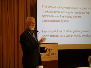
RozemaTed Rozema–Hans Seyle comments in his book, Calciphylaxis, about how PTH is a direct producer of calcium deposition in arterial walls.  My personal take on the deposition of calcium in arterial walls leading to atherosclerosis is that as we age, the fundus of the stomach does not produce the gastric acid needed to imbed a marker on the calcium molecule so it can be seen by the gut villi cells and be invited into the blood stream. This will cause not enough calcium to be absorbed to maintain the tightly controlled calcium balance in the blood stream. Over time, there is a miniscule parathyroid hormone release to take calcium from the bone to make up the shortfall.  This produces the sensitization to put some of the calcium into arterial walls.  Over many years, the clinical picture of early death and other vascular conditions result.

The issue of using calcium EDTA (instead of magnesium disodium EDTA to reduce metastatic calcium and treat vascular disease) is imbedded in the use of magnesium disodium EDTA.  Once the latter molecule hits the blood stream, the magnesium is dropped for chelation of calcium.  The resulting decrease in free serum calcium is what triggers the parathyroid action (and the platelet effect).  The resulting calcium disodium EDTA will then do all the toxic metal binding that calcium disodium EDTA does when given as a short IV infusion….

Joe Hickey—I believe that mercury is chelated with EDTA.  The resulting mercury EDTA rapidly vaporizes from urine and thus is difficult to measure.  If one believes that neither CaNa2EDTA or MgNa2EDTA effectively chelates mercury, then one must account for mercury with DMSA or DMPS.  If urine and fecal measurements are done, the largest amount of heavy metals removed is by far mercury with lead a distant second place.  If only urine is collected, lead is usually the highest excretion with mercury second, in my patients. Therefore, I also use DMSA if tolerable with either form of EDTA to account for mercury.  I will give 10 mg/kg of DMSA for three days, starting on the day of the IV with EDTA.  DMSA is less likely to vaporize in the stool. 

When treating primarily for vascular disease, I use MgNa2 EDTA 1.5 Gms over 1.5 hours with the addition of the DMSA.  I have found this regimen to be successful in patients who have had recurrent angina, post bypass, and stent closure.  I am rarely able to talk patients into consistently sitting for the 3 Gm/3 hour infusion.  I believe there is probably an additional effect for the 3 hour infusion in cases of calcific valvular disease and scleroderma, because of the parathyroid effect but I am not convinced that the 3 Gm dose is the sole treatment for vascular disease.  In younger patients with fibromyalgia or neuropathy, I will usually use CaNa2EDTA in combination with DMSA.  If DSMA is not tolerated, I will alternate with DMPS.

John Trowbridge— What are we trying to accomplish with chelation treatmenDr._John_Trowbridgets?  If a patient is substantially ‘loaded’ with toxic metals, treatment goals will be different than those for one with high-grade blockage disease in critical arteries. Patients suffering with crippling inflammatory and/or auto-immune diseases might require a another approach. 

Injectable and oral medication alternatives to EDTA should be considered, depending on a variety of factors.  However, 60 years of beneficial and safe reports regarding EDTA would argue for that to be the basic IV chelator.  Others could be added in ways that maximize their safety considerations as well, such as oral DMPS, DMSA, and d-penicillamine, as well as IM DFO.

With different stability constants and side effects, using two or more chelating agents at the same time, poses potential risks to the patient.  The simplest way to avoid such unpredictable interactions is to administer one chelating drug at a time. In 1993 I developed the idea of administering different chelating drugs on an intermittent, pulsatile schedule.  This protocol avoids the potential of interaction of two or more chelation drugs but still allows for exploiting the ‘preferences’ of each drug for different toxic metals.  Extra physiologic minerals would, of course, be needed to handle the additional chelator load.

To minimize side effects, I have patents take one 250 mg tablet each evening during the week.  For example, a patient might be advised to take d-Pen 250 mg each bedtime on days 1-7 of the month, then DMSA 250 mg each bedtime on days 8-14, then d-Pen again on days 15-21 and DMSA again on days 22-28, with a ‘break’ until day 1 of the next month.  I do not mix IV EDTA with DFO, nor do I give d-penicillamine  or DMSA within 4-6 hours of EDTA IV.  

With the changing urinary elements test reports, a patient might be told to take just d-Pen or DMSA every day of the month.  Now that oral DMPS is readily available, it might be inserted into the ‘weekly’ protocol as well, especially when mercury levels are substantially elevated on the test report.

By 1997, I had confirmed that our reduction of toxic heavy metal body burden – as measured by the d-Pen challenge urinary elements test – was proceeding at a rate at least one-third faster than with IV EDTA alone.

Two serious mistakes that beginning doctors often make when giving chelation therapy are insufficient attention to mineral depletion and too rapid reduction of cardiovascular medications.  Ample minerals must be supplied, either orally or IV, and gradual reductions of meds are required in order for the body to “relearn” more normal functions.         

Conrad Maudrmaulfairlfair—With calcium being an important factor in the aging process and in the atherosclerotic disease process, we want to have the maximum benefit from its removal that is possible.  I personally am a strong advocate of the 3-4 hour chelation treatment simply based on clinical experience.

Patients suffering from end-stage chronic degenerative diseases are misled when they read or are told that oral application of chelating agents and in particular EDTA can be an alternative to a comprehensive chelation therapy program including the parenteral administration of EDTA.  A patient having significant end-stage disease opting to take an oral chelation product because it is cheaper and does not require a personal commitment of time and involvement is most assuredly going to experience a poor outcome.  A practitioner who encourages a patient with significant disease to think that using oral chelation is better than doing nothing is being as irresponsible as allowing a patient uncontested to continue to smoke one pack of cigarettes a day because it is better than the two packs a day he/she had previously smoked.


TACT Trial: EDTA Chelation Therapy for Atherosclerosis

Effect of Disodium EDTA Chelation Regimen on Cardiovascular Events in Patients With Previous Myocardial Infarction

Lamas-Gervasio-CARDIOLOGYGervasio A. Lamas, MD; Christine Goertz, DC, PhD; Robin Boineau, MD, MA; Daniel B. Mark, MD, MPH; Theodore Rozema, MD; Richard L. Nahin, PhD, MPH; Lauren Lindblad, MS; Eldrin F. Lewis, MD, MPH; Jeanne Drisko, MD; Kerry L. Lee, PhD ;

Importance Chelation therapy with disodium EDTA has been used for more than 50 years to treat atherosclerosis without proof of efficacy.

Objective To determine if an EDTA-based chelation regimen reduces cardiovascular events.

Design, Setting, and Participants Double-blind, placebo-controlled, 2 × 2 factorial randomized trial enrolling 1708 patients aged 50 years or older who had experienced a myocardial infarction (MI) at least 6 weeks prior and had serum creatinine levels of 2.0 mg/dL or less. Participants were recruited at 134 US and Canadian sites. Enrollment began in September 2003 and follow-up took place until October 2011 (median, 55 months). Two hundred eighty-nine patients (17% of total; n=115 in the EDTA group and n=174 in the placebo group) withdrew consent during the trial.

Interventions Patients were randomized to receive 40 infusions of a 500-mL chelation solution (3 g of disodium EDTA, 7 g of ascorbate, B vitamins, electrolytes, procaine, and heparin) (n=839) vs placebo (n=869) and an oral vitamin-mineral regimen vs an oral placebo. Infusions were administered weekly for 30 weeks, followed by 10 infusions 2 to 8 weeks apart. Fifteen percent discontinued infusions (n=38 [16%] in the chelation group and n=41 [15%] in the placebo group) because of adverse events.

Main Outcome Measures The prespecified primary end point was a composite of total mortality, recurrent MI, stroke, coronary revascularization, or hospitalization for angina. This report describes the intention-to-treat comparison of EDTA chelation vs placebo. To account for multiple interim analyses, the significance threshold required at the final analysis was P = .036.

Results Qualifying previous MIs occurred a median of 4.6 years before enrollment. Median age was 65 years, 18% were female, 9% were nonwhite, and 31% were diabetic. The primary end point occurred in 222 (26%) of the chelation group and 261 (30%) of the placebo group (hazard ratio [HR], 0.82 [95% CI, 0.69-0.99]; P = .035). There was no effect on total mortality (chelation: 87 deaths [10%]; placebo, 93 deaths [11%]; HR, 0.93 [95% CI, 0.70-1.25]; P = .64), but the study was not powered for this comparison. The effect of EDTA chelation on the components of the primary end point other than death was of similar magnitude as its overall effect (MI: chelation, 6%; placebo, 8%; HR, 0.77 [95% CI, 0.54-1.11]; stroke: chelation, 1.2%; placebo, 1.5%; HR, 0.77 [95% CI, 0.34-1.76]; coronary revascularization: chelation, 15%; placebo, 18%; HR, 0.81 [95% CI, 0.64-1.02]; hospitalization for angina: chelation, 1.6%; placebo, 2.1%; HR, 0.72 [95% CI, 0.35-1.47]). Sensitivity analyses examining the effect of patient dropout and treatment adherence did not alter the results.

Conclusions and Relevance Among stable patients with a history of MI, use of an intravenous chelation regimen with disodium EDTA, compared with placebo, modestly reduced the risk of adverse cardiovascular outcomes, many of which were revascularization procedures. These results provide evidence to guide further research but are not sufficient to support the routine use of chelation therapy for treatment of patients who have had an MI.

Trial Registration clinicaltrials.gov Identifier: NCT00044213

Treatment of lead toxicity with chelation was first reported with EDTA in the early 1950s.1Apparent success in reducing metastatic calcium deposits2 led Clarke et al3 in 1956 to treat angina patients with EDTA, and others to use chelation for various forms of atherosclerotic disease.4– 6Chelation therapy evolved to constitute infusions of vitamins and disodium EDTA, a drug that binds divalent and some trivalent cations, including calcium, magnesium, lead, cadmium, zinc, iron, aluminum, and copper, facilitating their urinary excretion.7,8

Over the next decades, based on favorable anecdotal and case report experience, chelation practitioners increased their use of EDTA for coronary and peripheral artery disease. The 2007 National Health Statistics Report compared chelation use since 2002 and noted an increase of 68%, from 66 000 to 111 000 adults using chelation therapy,9 although the indications for therapy were not clearly defined, and the prevalence of use of chelation therapy for cardiovascular disease is unknown.

Three small clinical trials have assessed the effects of chelation on surrogate outcomes, such as walking distance in patients with claudication (2 trials with 185 patients total) and time to exercise-induced ischemia in patients with coronary disease (1 trial with 84 patients). These studies did not find any evidence of treatment efficacy but were underpowered for evaluation of clinical events.10– 12 As a consequence, mainstream medical organizations consider the therapeutic value of chelation for atherosclerotic vascular disease unproven13 and the use of this therapy potentially dangerous. Disodium EDTA, particularly when infused too rapidly, may cause hypocalcemia and death.14 The Trial to Assess Chelation Therapy (TACT) was conducted to respond to the public health problem posed by EDTA chelation therapy: large numbers of patients being exposed to undefined risks for unproven benefits.

http://jama.jamanetwork.com/article.aspx?articleid=1672238

Chelation Therapy for Peripheral Arterial Occlusive Disease

blood-1237550-639x450E. Ernst, MD, PhD, FRCP (Edin)

Abstract:

Chelation therapy is viewed, promoted, and practiced as a form of complementary/alternative medicine (see References 1 and 21 2 ). It uses repeated intravenous administration of EDTA, usually in combination with vitamins, trace elements, and iron supplements as a treatment for a variety of diseases.3 4 5 Oral chelation therapy also has been advocated (see Reference 66 ) but will be excluded from this review. Among others, chelation therapy is claimed to be effective in reversing the arteriosclerotic disease process,7 8 in particular peripheral arterial occlusive disease (PAOD). Proponents claim that it can be an alternative for amputation in severe cases.8 This goes back to an observation made in the 1950s, when it was noted that patients undergoing EDTA therapy for lead poisoning felt a relief of angina pectoris after this therapy.9 While various authors9 10 11 12 13 1415 have subsequently reported promising results in uncontrolled studies, others have been unable to confirm these in similarly designed trials.16 17 The debate of whether or not it is a useful form of therapy for PAOD therefore continues.18 19 2021 22 23 24 25

Meanwhile, numerous clinics have been set up in Europe, the United States, and elsewhere using and promoting chelation therapy for PAOD. In the light of recent trial data, it is therefore timely to systematically review the evidence from randomized, placebo-controlled, double-blind trials and ask whether chelation therapy for PAOD is safe and effective.

http://circ.ahajournals.org/content/96/3/1031.long


EDTA chelation therapy in the treatment of vascular disease.

Chappell LT1, Janson M.

Abstract

A retrospective analysis of treatment results from 2870 patients, with various chronic degenerative and age-associated diseases, who were treated with di-sodium magnesium EDTA chelation therapy, suggests that the case against EDTA Chelation Therapy should be re-opened.

Using qualitative but never-the-less standardized criteria for improvement, our analysis shows that EDTA Chelation Therapy resulted in “marked” improvement in 76.89% and “good” improvement in 16.56% of patients with ischemic heart disease; also, “marked” improvement in 91% and “good” improvement in 7.6% of patients with peripheral vascular disease and intermittent claudication. In a group of patients with cerebro-vascular and other degenerative cerebral diseases, 24% had “marked” improvement, and 30% had “good” improvement. Of four patients with scleroderma, three had “marked” improvement and one had “good” improvement. Seventy-five percent of all of the patients had “marked” improvement in “geriatric symptomatology of vascular origin”.

The authors recommend renewed study of EDTA Chelation Therapy. The possibility of a “tomato effect”, i.e., a drug which works, but the majority of physicians believe that it doesn’t work, needs to be ruled out. A favorable climate needs to be created, in which FDA-approved studies of its usefulness in treating peripheral vascular disease can take place.

http://www.ncbi.nlm.nih.gov/pubmed/8820322

Should EDTA chelation therapy be used instead of long-term clopidogrel plus aspirin to treat patients at risk from drug-eluting stents?

by Chappell LT, 2007red-pills-1526972-639x384

Abstract: The recently discovered increased risk of blood clots, leading to myocardial infarction and sudden death beginning six months after medicated stents are implanted in patients following percutaneous transluminal coronary angioplasty (PTCA), has left cardiologists pondering what course of action to take. The purpose of adding implanted medication to a stent is to prevent thrombin accumulation and restenosis. However, these stents may increase, rather than decrease, the risk. Although long-term treatment with clopidogrel bisulfate (Plavix) plus aspirin for at least 12 months has been suggested as a preventive treatment, there is no evidence from randomized, controlled trials that this treatment is effective for more than six months. Clopidogrel also increases the risk of major bleeding episodes. The author served as the primary investigator for a study that showed cardiovascular patients treated with EDTA chelation therapy had a lower rate of subsequent cardiac events, including myocardial infarction and death, than those treated with cardiac medications, PTCA, or coronary artery bypass graft (CABG). The data also indicated chelation therapy might be effective in preventing thrombosis and cardiac events from stent implantation. There is evidence EDTA chelation therapy might prevent hypercoagulability resulting from the placement of stents, although not specifically medicated stents. Based on the limited data currently available, intravenous EDTA may be safe and effective for treating patients who have implanted medicated stents. Prospective clinical trials are needed, and EDTA should be included in those trials.

http://www.ncbi.nlm.nih.gov/pubmed/17604460

Chelation Therapy: Clinically Relevant or Just Quackery?

ronhoffmanRon Hoffman, MD; Feb 10, 2014

Chelation therapy, a type of intravenous (IV) treatment promoted by some members of the complementary and alternative medicine community, has long been mired in controversy.

Often dismissed as quackery, chelation therapy was the subject of a recently completed NIH study (Trial to Assess Chelation Therapy [TACT]) that showed the practice to be of moderate benefit to heart-attack survivors. Yet the controversy continues unabated, with some calling the study misguided or flawed and few in the conventional medical community willing to embrace chelation therapy as a legitimate option for heart patients.

How Chelation Therapy Works

Chelation therapy consists of a series of IV administrations of disodium or calcium ethylenediaminetetraacetic acid (EDTA) mixed with minerals and vitamins.

Typically, patients recline in a chair in the clinician’s office for one to four hours once to three times weekly for a series of 20 to 80 chelations. “Booster” chelations may be administered on a monthly basis or intermittently for years.

The purported benefits of chelation therapy vary but typically include the following:

  • Improves such circulatory disorders as coronary artery disease (CAD), cerebrovascular disease, or peripheral vascular disease;
  • Detoxifies the body of such heavy metals as lead, cadmium, and mercury; and
  • Combats degenerative diseases and slows the aging process.

The procedure is usually not covered by Medicare or private insurance. The out-of-pocket costs are borne by patients and may total thousands of dollars for a single course of treatment.

History

The term chelation (derived from the Greek chelos or claw) refers to the mineral- or metal-binding properties of certain compounds that can hold a central cation in a pincerlike grip. Developed in Germany in 1935, EDTA was originally used as a means of binding and extracting calcium in the dye industry.

In the 1940s, Martin Rubin, Professor Emeritus of Biochemistry at Georgetown University Medical Center in Washington, D.C., discovered EDTA’s effects on calcium in biological systems. This discovery led to the product’s use as an anticoagulant and is still used in “purple top” blood-collection tubes. Professor Rubin’s research led him to advance the use of EDTA for treatment of hypercalcemia and, eventually, lead poisoning.

In the 1950s and 1960s, several clinicians began to observe that patients treated for lead poisoning with IV EDTA experienced improvements in their cardiovascular conditions. This observation led to the widespread, but mostly empirical, use of EDTA therapy for heart patients within a growing community of alternative medicine practitioners.

Studies were undertaken, but these were mostly observational or uncontrolled and involved only small numbers of patients. Chelation therapy for other than the approved indications of refractory hypercalcemia or severe lead toxicity remained highly touted but poorly substantiated.

Clinicians practicing chelation therapy were sometimes targeted by medical boards for disciplinary action, irrespective of whether specific patient harm had occurred. Some states adopted regulations prohibiting the practice of chelation therapy. To this day, disodium EDTA is not approved by the FDA to treat any diseases. However, disodium EDTA is produced by compounding pharmacies for individual patients, so the treatment can still be obtained.

In 1998, the U.S. Federal Trade Commission (FTC) targeted the American College for Advancement of Medicine (ACAM), an organization that has trained and certified physicians in methods of safe administration of chelation therapy since the 1970s, for allegedly outsized advertising claims made regarding the treatment of atherosclerosis.

The FTC concluded that there was a lack of scientific studies to support these claims and that pro-chelation statements made by ACAM were false. As an alternative to litigation, ACAM stipulated that it would curtail public pronouncements presenting chelation therapy as an effective treatment for heart disease.

The public’s enthusiasm for chelation therapy remained undiminished, however. Between 2002 and 2007, use of chelation therapy to treat heart disease and other conditions grew in the United States by nearly 68% to 111,000 people.1 As of the start of the TACT Study in 2001, it was estimated that patients received 800,000 individual EDTA infusions per year.2

Until the TACT study, mainstream clinicians widely believed that EDTA chelation therapy for conditions other than acute lead intoxication was an unwarranted and dangerous modality. This is true to the extent that excessive doses of EDTA can be nephrotoxic; cases of renal failure resulting in dialysis or death have been recorded. Additionally, transient hypocalcemia provoked by EDTA calcium sequestration can trigger cardiac arrhythmias or sudden death.

But these outcomes have generally occurred only in rare instances where EDTA is administered in too high a dose and/or too rapidly or without regard to a patient’s glomerular flow rate. In 1989, a “Protocol for the Safe and Effective Administration of EDTA” was developed and subsequently updated.3

The detailed protocol provides strict criteria for patient selection and cautions clinicians to perform an initial evaluation of renal function using the Cockcroft-Gault equation and to frequently monitor renal function throughout a series of chelation treatments. Emergency procedures are outlined should adverse reactions occur.


Read More:

http://www.empr.com/chelation-therapy-clinically-relevant-or-just-quackery/article/333324/

Report on the Proceedings of a Summit on New Directions for Chelation Therapy

Participants in the Summit for New Direction in Chelation Therapy

From March 13 to 15, 2013, the International College of Integrative Medicine (ICIM) held a summit meeting about what should be accomplished next, now that EDTA chelation therapy has been supported as a useful treatment for vascular disease by the Trial to Assess Chelation Therapy (TACT).  Experts from around the world were invited.  This paper is a summary of the conclusions and recommendations of this gathering.  Key presentations were given by Drs. John Trowbridge, Efrain Olzsewer, and Eleonore Blaurock-Busch.  Representatives from the U.S., Canada, Indonesia, Brazil, Denmark, the Netherlands, Germany, Ecuador, and New Zealand participated, as well as the attendees for the Advanced Metals Workshop that was part of the spring meeting of ICIM.  Recordings of the lectures are available from icimed.com. This paper was prepared prior to articles on TACT published in the Journal of the American Medical Association in the March 27, 2013 issue.

Background

EDTA has been used as a treatment for vascular disease since Norman Clarke, Jr.’s work in 1952.  For a timeline of the many studies that have supported its effectiveness, see chelation.me.  In 1981, the AMA challenged the proponents of chelation therapy to produce a large-scale, randomized, controlled, clinical trial to prove its safety and effectiveness.  The members of the American College of Advancement in Medicine (ACAM) led by president Ross Gordon collaborated with Walter Reed Hospital to begin such a study for treatment of peripheral vascular disease in 1987.  Unfortunately, the first Gulf War took the investigators away from the study, and it was not completed.  In 1999, Congressman Dan Burton, Chair of the Committee on Oversight, held a hearing bringing together the head of the Heart, Lung, and Blood section of the National Institutes of Health and several physicians who testified about their experiences with chelation.  NIH subsequently called for proposals, and eventually TACT was funded, with Gervasio Lamas as chief investigator.

TACT was unique in that it combined university research cardiologists and experienced chelation specialists with private offices.  134 sites from the U.S. and Canada participated in the randomized, placebo-controlled, double-blind, clinical trial.  TACT continued for 7 years, and included 1708 patients with documented previous heart attacks who continued to receive evidence-based therapy.  The primary end point was a composite of new cardiac events to include death, heart attack, stroke, hospitalization for unstable angina, and need for revascularization surgery.  TACT showed that the therapy was unquestionably safe, and the group treated with chelation therapy had fewer cardiac events, which was statistically significant.  The results were announced by Lamas at the American Heart Association meeting on November 4, 2012 in Los Angeles.  Publication of the results is pending.  The authors called for further studies to confirm the results and explore the mechanisms of action.

Where we stand now, according to the Summit

  1. TACT conclusively showed that chelation therapy used according to the recommended protocol is safe.
  2. TACT and the many other studies that proceeded it support the use of chelation therapy as an option for patients with vascular disease, especially for those who also have diabetes and those with a history of anterior wall myocardial infarction.
  3. There is not yet enough evidence to state that chelation therapy should be given to all cardiac patients.  More studies need to be done.  A duplication of TACT would be ideal, as long as it included heavy metal testing.  However, another $30 million to repeat the study might be difficult to find.
  4. Strong consideration should be given to doing a challenge test for heavy metals (especially lead) for all patients with vascular disease.  If high levels are found, the patients should be treated with chelating agents.
  5. Regulatory agencies, such as medical boards, should immediately stop harassing physicians who offer chelation therapy to their patients who give appropriate informed consent.  Physicians who offer chelation therapy have accomplished exactly what the AMA asked them to do in 1981 to justify its use.
  6. Most physicians who offer chelation therapy are happy to serve as consultants for placebo-controlled RCT’s, but are uncomfortable with the ethics of giving placebos to patients who have come to them for help.  Certainly, patients should not be asked to pay to receive placebos, especially for a potentially life-threatening illness.  Physicians who provide chelation are almost always convinced that in their experience the therapy is very effective.
  7. Many chelation doctors feel that their primary goals of showing efficacy and safety with a RCT have been accomplished with TACT.  Gaining FDA approval of EDTA for use in vascular disease is secondary, and they encourage qualified investigators to move in that direction.

Recommendations of the Summit

  1. More research should indeed be done on metal toxicity, free radical pathology, and on various diseases that have been linked to free radical pathology, especially vascular disease.
  2. Chelation doctors do not have the resources to fund or carry out clinical trials, but they do have the expertise to help plan them.
  3. The conditions that are most likely to show benefit with chelation treatment and thus should have the greatest research priority are as follows:
    1. Patients waiting to have limbs amputated due to non-infected vascular disease.  For end points, all that is needed is to count the remaining limbs.  Claus Hancke’s work is most impressive in this regard.

b. Walking distance and A/B index in patients with peripheral vascular disease.  In our experience, a very high percentage of patients improve.  Ffrain Olzsewer and Jim Carter documented this.  There have been a couple of negative studies published on this subject in prominent journals, but they have been seriously flawed. Stephen  Olmstead has written a good research protocol to evaluate chelation treatment for peripheral artery disease that is almost ready to go.  He is willing to share his work with others. Attendees to the summit expressed significant concern that opponents of the therapy might proceed with new studies that are designed to fail, which has happened in the past.

  1. Brachial artery stiffness and other measurements of vulnerable plaque.  Peter van der Schaar is beginning a study on arterial stiffness.
  2. Diabetic patients who have evidence of vascular disease.
  3. Patients who have suffered an anterior wall MI.
  4. Patients who have angina that is difficult to control with drugs.
  5. Macular degeneration.
  6. Patients who have been told that revascularization surgery is an option
  7. Quality of Life measurements should be included in all research projects.  Chelating physicians insist that their patients feel considerably better with long-term treatment, even though the relatively short follow-up in TACT detect significant improvement.
  8. Other areas that are important to study and are likely to show successful outcomes:
    1. Patients with hypertension and elevated lead levels
    2. Arterial intimal thickness and high resolution ultrasound of the carotid arteries (see the work of Robert Bard)
    3. Osteoporosis
    4. Mild to moderate Alzheimer’s disease associated with heavy metal toxicity
    5. Autoimmune diseases, especially scleroderma
    6. Fibromyalgia with high levels of toxic metals detected with a challenge test
    7. Diseases that are familiar to the public should be studied in order to raise awareness and support for chelation.
  9. There are many biomarkers in the laboratory that can help examine the mechanisms of action of chelation therapy.  Expert biochemists (Blaurock-Busch, Jaffe, Quig) are happy to consult with investigators as to which ones are most appropriate to utilize in this assessment.
  10. Various combinations of chelating agents, and different doses of such entities as EDTA and vitamin C are important to study.
  11. Chelation therapy is useful to study at all stages, to include

a.     Preventive

b.     Pre-emptive (early signs of disease)

c.     Treatment of established disease

d.     Treatment following revascularization  procedures

e.     Maintenance treatments are very important

  1. Such international lecturers as van der Schaar, Olzsewer, Rozema, Hancke, Dooley, and Godfrey continue to teach physicians on how to use chelation therapy safely and effectively.  Organizations such as ACAM, ICIM, and A4M hold workshops in the United States.  Excellent recent textbooks have been published by van der Schaar and Blaurock-Busch (both are available through the International Board of Clinical Metal Toxicology).  There is a need to move toward consistent protocols and best practices.
  2. Studies must be well-designed and conducted so that clear outcomes can be readily understood and will resonate with a large portion of the population, as well as stimulate Congressional action.
  3. Use of NBMI—a compound being studied by Boyd Haley might turn out

to be a powerful therapeutic modality.

Conclusion

Raising public, political, and media awareness is now essential.  Experienced chelating physicians can help provide solid data to support general understanding of efficacy, mechanisms, and positive outcomes in the treatment of vascular diseases. Registries might be the best way for clinicians to collect data without the constraints of a RCT.  Self-insured corporations, such as Parker-Hannifin are now paying for chelation therapy.  Cooperation among organizations with similar interests, such as ICIM, ACAM, AAEM A4M, ABCMT, IBCMT, and specialized laboratories is strongly encouraged to standardize protocols and set up registries.  This can be done quickly and with minimal expense.  Physicians from around the world should be included.  Experienced chelating physicians can serve as consultants for researchers who are qualified to perform RCTs.  NIH and various foundations are encouraged to fund projects discussed in this paper.  Pollution with heavy metals continues to get worse, and evidence is mounting that their toxicity is an important factor in the development of chronic degenerative diseases.

Chappell: Saving a Million Hearts

Saving a Million Hearts workshop at ICIM

By L. Terry Chappell

Introduction

In September of 2011, the Department of Health and Human Services (DHHS), the Centers for Disease Control(CDC), and the Centers for Medicare and Medicaid Services(CMS)  jointly announced the Million Hearts Initiative(1,2).  The goal is to prevent 1 million heart attacks and strokes over the next five years.  Other groups such as the American Heart Association, the American College of Cardiology, the American Pharmacy Association, and Walgreen drug stores quickly joined the effort.

Unfortunately, the action plan to achieve this lofty goal as published is likely to fail.  Nevertheless, those of us in Integrative Medicine should embrace the overall goal and use all of our skills to formulate a plan to prevent even more heart attacks and strokes than the efforts put forth by these prestigious organizations.  This article analyzes the strengths and weaknesses of the Million Hearts Initiative(MHI) and shows how we can make it dramatically better.

Cardiovascular Disease as the Leading Cause of Death

In the United States there are about 2 million heart attacks and strokes each year with 800,000 fatalities.  Not only is this the leading cause of death, but also the overall medical cost of these diseases is estimated to be $450 billion per year.  From 1980 to 2000 there was a significant reduction in the death rate from cardiovascular disease, most of which was due to lifestyle changes and preventive medicine.  Yet cardiovascular disease is still by far the leading cause of death.  HHS Secretary, Kathleen Sebelius, states that heart disease is responsible for one of every three deaths in the U.S.

The MHI Plan to Prevent Heart Attacks and Strokes

The clinical interventions put forth by the MHI consist of four potential categories of drugs.  The treatment acronym is the ABC’S of prevention:  aspirin for high-risk patients, medications to control blood pressure, cholesterol management, and smoking cessation if needed (varenicline, nicotine patches, etc.).  In addition, the MHI calls for improved nutrition through a reduction in the intake of sodium and trans-fats.  The MHI hopes to coordinate activities with Obama’s Affordable Care Act.  Electronic health records and quality recognition programs offered by both the government and various private insurance plans should also be useful for recruitment of patients to participate.

At present, only 47% of patients at risk take aspirin, 46% have blood pressure under control, and 33% have LDLs below 100.  The specific goals of the MHI are to increase all of these numbers to 65% by 2017.  A fourth clinical goal is to reduce smoking prevalence from 19% to 17%(1).

Emphasizing four interventions that might require drug therapy certainly makes one wonder about the influence of the pharmaceutical industry in this effort. There are at least 30 million people in the U.S. whose blood pressure and/or cholesterol are not under control.  That is a pretty large target population, just with these two factors.  Overall, Forbes estimates that the MHI seeks to put half of our adult population on drugs prescribed by doctors.

One of the strengths of the MHI plan is that it does not depend on intensive care by cardiologists and vascular surgeons.  In fact, several popular blogs written by these specialists have complained that cardiologists are being left out of the campaign.  Perhaps there is a reason for this omission.  The OAT trial(3) in 2006 demonstrated that opening totally occluded arteries with stents after uncomplicated myocardial infarctions involving those vessels actually increased the mortality rate when compared to medical management. Soon afterwards, the COURAGE trial(4) showed that angioplasty and stents for stable coronary artery disease were no more effective than proper medical management.  Before the COURAGE trial, 85% of all stents in the U.S. were surgically placed in patients with stable coronary artery disease. Both of these important studies have been virtually ignored in clinical practice.  Vascular specialists continue to place unnecessary stents in many patients each year.  A recent JAMA editorial(5) described this practice as an “expensive placebo”.   The authors further commented that “some entire medical subspecialties (might be) based on little evidence”.  No doubt there are valid indications for revascularization procedures and complex drug therapy.  Cardiologists are necessary.  Many of them would be more effective, however, if they focused more on nutritional biochemistry.

Of great interest is the study by Canto and associates(6) that analyzed 542,008 patients who had heart attacks from 1994 to 2006.  For those patients who suffered their first heart attack, the in-hospital mortality was inversely proportional to the number of traditional risk factors that were identified.  The risk factors they examined were hypertension, smoking, dyslipidemia, diabetes, and family history of heart disease.  Obviously, other factors were contributing to the increased mortality for these patients.  If we are to succeed, we must do a more thorough job of identifying risk factors and modifying them safely.

Criticism of the MHI Plan

The most obvious deficit in the MHI plan is that it does not include three commonly recognized lifestyle factors for the prevention of cardiovascular disease: regular exercise, stress coping measures, and weight reduction if needed.  Exercise alone is probably more effective than any drug one can take.  By excluding these important lifestyle factors it becomes highly unlikely that the MHI will succeed in real life.

The MHI appropriately states that we must reduce trans-fats and sodium in our diets, but it could do much more.  At the very least, patients at risk should avoid foods that are high in the glycemic index, aspartame, high-fructose corn syrup, processed foods, and fried foods.  We also could eat organic raw veggies as much as possible.  The use of unrefined salt would add beneficial trace minerals.  Not surprisingly, the potential benefits of nutritional and herbal supplements are not mentioned in the MHI.

Diabetes is another prominent risk factor for cardiovascular disease.  Weight control and low carbohydrate diets are important for prevention and treatment of diabetes.  Diet, exercise and supplements are often sufficient to achieve control of Type 2 disease without medications.

Poverty and inequality are factors that have been shown to increase cardiovascular disease.  Not only do these factors cause economic stress, but they also result in poor quality food and increased smoking as a stress-coping measure.  Such socioeconomic factors make it more difficult for the ABC’S of the MHI to succeed.  A more comprehensive approach as I describe is required to overcome the twin risk factors of poverty and inequality.

The MHI is careful to note that aspirin and statin drugs for cholesterol management are to be used only for high-risk patients.  However, that might serve to be the “fine print” that nobody reads.  Recent reports show that for primary prevention of cardiovascular disease the “number needed to treat” to prevent one heart attack with aspirin is 163 and for statin drugs is 200(7).  The “number needed to harm” for both of these interventions is much lower.  Thus the use of these drugs for primary prevention is highly questionable.  However, many physicians still prescribe them when not indicated, which is a waste of resources and the potential source of serious complications.

If we are going to succeed in saving a million hearts with our current socioeconomic and lifestyle stresses and our failure to change our therapies in response to definitive evidence, we should look at additional risk factors, especially ones whose remedies are much less likely to cause complications than the proposed drugs.  We should emphasize powerful lifestyle changes and safe, optimal supplements instead of diverting our attention toward aspirin, anti-hypertensive drugs, and statins. For this, we can rely on and offer our patients the insights and experience of integrative medicine.

Let’s Get Serious About Saving a Million Hearts

Obviously, we cannot save a million hearts and strokes all by ourselves.  But we can save way more than our share.  First, we should identify the hearts that need saving (although the case can be made that all hearts need saving).  We can determine if patients’ hearts are at risk mostly by performing a history and physical exam and gathering basic lab and other tests, some of which might have been previously been performed.  If patients have a history of documented vascular disease, hypertension, hyperlipidemia, diabetes, smoking in the previous 5 years, or a family history of heart attacks or strokes, they automatically qualify.

Computerized risk assessments, usually based on the Framingham Risk Assessment, might or might not be helpful.  They provide striking graphic displays that demonstrate the effect of improving basic risk factors.  However, they don’t include the cumulative effect of a comprehensive risk factor plan like we are discussing.  If patients are at least 50 years old or the physician suspects high-risk lifestyles, one or more screening tests to determine if they are beginning to develop plaque in their arteries is indicated.  If a resting EKG has non-specific ST/T-wave changes, their heart might be at risk.  A stress EKG can have false positives and false negatives, especially in women.  A stress echocardiogram is more accurate in females.  An ultra-fast CT scan for calcium score is a good screening test.  A carotid intima media thickness(CIMT) ultrasound test by CardioRisk(www.cardiorisk.us) is also a very sensitive screening test that can be done by that company periodically in your office.  The ankle/brachial index is a reasonable screen for peripheral artery disease, although not very sensitive, in my experience.  If positive, however, there is an increased risk for heart attacks and strokes.

If we determine that a patient is at risk, a comprehensive cardiovascular risk factor evaluation is indicated.  For our patients who join the MHI, we often recommend a VAP cholesterol panel, including Lp(a)(8), HbA1C, ferritin, fibrinogen, CRPsensitive, red cell magnesium, 25 [OH] vitamin D3(9), and homocysteine test.  Virtually all of these tests and more are included in a comprehensive cardiovascular blood panel.  Two companies that offer such panels are Doctors Data(www.doctorsdata.com) and Atherotech(www.Atherotech.com).  We also do a EDTA challenge test for heavy metals, with special attention to lead(10).  If available, heart rate variability testing frequently detects high sympathetic activity that is not balanced by parasympathetic output, even when the patient is unaware of excessive stress.   A saliva test strip for nitric oxide (www.advancedbionutritionals.com) can detect low NO levels, which theoretically at least, can be improved with nutritional support.  Other tests for nutritional factors can certainly be ordered, but they are beyond the scope of this article.

In our report of findings, we estimate how much risk we think each patient has and how we feel we can improve that risk with various interventions.  Our individual patient data base is considerably larger than that of the MHI.  Our recommended treatment interventions include more aggressive lifestyle measures, nutritional supplements, herbal therapies, and other treatments as indicated.

Integrative Treatment Plan

Start with the ABC’S.  Instead of or in addition to aspirin, to reduce platelet aggregation, we can use fish oils, garlic, vitamin E (mixed tocopherols especially gamma), nattokinase, and/or lumbokinase.  Donating blood several times a year is another way to decrease blood viscosity.  One study showed an 88% reduction in the risk for myocardial infarction for 153 middle-aged men who donated blood in the previous 24 months(11)  That study has been criticized, but a more recent study(12) delineated a more complex mechanism and confirmed that blood donation might reduce the risk of vascular disease.  In addition to reduced blood viscosity, the resulting decrease in elevated ferritins substantially lowered free radical activity.  Rheologics (610-524-5427) makes a machine that measures blood viscosity.

The blood pressure might respond to garlic, potassium, magnesium, and other phytonutrients.  I have found rauwolfia with sandalwood and other herbs(BP Natural Relief) to be particularly effective(www.natrelief.com).  Weight loss can often lower the blood pressure significantly. These measures might be sufficient by themselves, or they can be used in conjunction with medications to achieve good control.

For cholesterol, HDL, and LDL management, low carbs appears to be the most effective diet(13), especially if the triglycerides are high.  But this remains controversial.  The DASH, LEARN, Ornish and Mediterranean diets are alternatives.  Red yeast is a natural statin that can effectively lower cholesterol and LDL, with much fewer side effects than the drugs.  As with statin drugs, the main beneficial effect from red yeast rice might be to reduce arterial inflammation rather than to reduce LDL.  Always replace coenzymeQ10 when prescribing any kind of statin.  Both muscle inflammation and congestive heart failure have been attributed to low levels of coQ10, which is depleted by the statins.  Fish oils can help reduce cholesterol and so can cinnamon, niacin, berberine, and lecithin.  Intravenous essential phospholipids from lecithin have been used in Europe to treat coronary artery disease.  Proteolytic enzymes might also be effective to reduce inflammation.  Food allergies can be important, especially gluten and casein sensitivity.  A therapeutic trial of an elimination diet can be very helpful.

To stop smoking, hypnosis and acupuncture are somewhat effective. The medication varenicline(Chantix) might have its place, but the incidence of side effects is troubling.

For better fitness compliance an exercise prescription is mandatory, depending on the physical capacities of patients.  People often need to have specific goals to get the best results.  Al Sears’ PACE program with brief periods of intense exercise makes sense to me.  It is backed by the Harvard Professional Lifestyle Study(14).  Adequate fitness, however, can usually be achieved by walking for 30 minutes 5 days per week.

Always be aware of how important stress can be for cardiovascular disease.

One of the best-documented treatment programs is Heart Math(15), which is a home tutorial using biofeedback.  Yoga, meditation, progressive relaxation, visualization, deep breathing, emotional freedom technique, prayer and acupressure are procedures that can be utilized.  All patients in the MHI should form a plan to improve their stress-coping activities, especially if their heart rate variability results are abnormal.

Nutrient deficiencies are frequently detected with the comprehensive cardiovascular risk profile, particularly magnesium.  Antioxidants are indicated if an increased amount of oxidized LDL is detected.  Linus Pauling’s admonition to treat patients who have elevated Lp(a) levels with vitamin C, proline, and lysine still rings true.  The optimal level of 25 [OH] Vitamin D3 is 60-100 ng/ml, although the listed normal is usually as low as 30 ng/ml.  Calcium might be given to lower the risk of osteoporosis or colon cancer, but always balance it with at least half of the milligram dose of magnesium.  Do not prescribe the ultra-high doses of 1500-2000 mg of calcium a day.  Studies have shown that high-dose calcium can lead to calcification of the arteries.  Coenzyme Q10, d-ribose, and l-carnitine are helpful adjuncts, especially for congestive heart failure and fatigue.  Medium chain triglycerides from coconut oil are useful to preserve brain function.  The herb, apoaequorin(Prevagen) is particularly good to preserve memory, in my experience.  The physician formulation of apoaequorin is four times as strong as the product available over-the-counter.

For many years, integrative physicians have found intravenous EDTA chelation therapy to be very effective in treating and preventing cardiovascular disease.  This is especially true if a build-up of toxic metals is detected.  Lead is the best-documented toxic heavy metal(10).  It has been linked to heart disease, cancer and autoimmune problems.  If mercury is found, DMPS or DMSA might be needed in addition to EDTA.  The published intravenous EDTA protocol appears to be effective, even if heavy metals are not found.  The author and associates demonstrated a dramatic decrease in subsequent cardiac events in high-risk patients who had received chelation therapy(16).  The results of the Trial to Assess Chelation Therapy (TACT) are due to be published this summer.

An under-appreciated advantage of enrolling a patient in a course of chelation therapy is that the treatments are given once or twice a week during the basic course.  That means that each week, the nurse has a teaching opportunity to reinforce diet, exercise, stress-coping, supplement compliance, and habit control, all of which are important for saving hearts.  Our staff helps the patient set goals and identify barriers to reaching the goals.  As with any class or program, repetition is key.  It often helps to bring a friend.  When patients share their experiences and goals with others, results can be better than trying to follow the program by themselves.  Group visits to deal with risk factors and lifestyle might be a useful service to offer.

Monitoring and maintenance are two key concepts for a successful program.  The risk factors identified must be monitored often enough to assure that interventions are effective.  Too often the patient and the physician identify risk factors, correct them temporarily, but fail to be sure that the factors remain under control.  Non-invasive vascular tests should be repeated to monitor progress.  Lab biomarkers should be repeated at specified intervals.  The CIMT and the heart rate variability are particularly good monitoring tests.  However, the ultra-fast CT scan is not.

A summary of the integrative approach in seven steps is outlined in Table 1.

Research and New Frontiers

Several avenues of research are currently taking place, including genomics, molecular targeting, stem cell biology, and regenerative medicine(17).  Both conventional and integrative medicine are active in these areas of interest.  Progress is anticipated within the five-year target period of the MHI.  For example, stem cells harvested from autologous bone marrow are being tested to treat myocardial infarction(18).  Initial results were not impressive, but the authors were optimistic that revisions in protocol might yield better results.  Mikirova and associates(19) recently showed that chelation of heavy metals improved the number of stem/progenitor cells in circulation.  Our version of the MHI should be a fluid plan that can be improved as new evidence emerges.

One criticism of integrative medicine is that there are few large clinical trials to support the therapies that are utilized.  Harvard professors Groopman and Hartzband in their book, Your Medical Mind (7), point out that too often the larger the clinical trial, the less significant the results.  Their reason is that it takes a large study to have sufficient statistical significance to prove a minimal effect.  Smaller studies with larger effects are often more useful.

On March 31, 2012 in Lexington, Kentucky, the International College of Integrative Medicine will hold a forum on the Million Hearts Initiative for clinicians experienced in the use of chelation therapy and other integrative techniques.  Round table discussions by the experts will explore further the ideas presented in this article.  Readers are invited to attend.  The proceedings will be published in the Townsend Letter.

Conclusion

How much effort is required to prevent a heart attack or a stroke?  How about a million heart attacks and strokes?  We applaud the conventional medical community and government for setting the MHI as a lofty goal.  Unfortunately, it is unlikely that goal will be reached with the plan that has been put forth.  On the other hand, utilizing a comprehensive, integrative approach, we can make a huge impact for those one million individual hearts and brains that we want to save.  Not infrequently, hypertension and hyperlipidemia can be controlled by detoxification of heavy metals, exercise, a healthy diet and stress management without the use of medications that might cause more adverse affects than beneficial ones.  Nutritional and herbal supplements, as needed, can be added with greater safety than many medications, with similar benefits.

Patients must be presented with all the evidence in an unbiased manner.  Then it is their responsibility to choose the therapies that suit them best. Individual treatment plans are more effective than rigid guidelines.  Our goal is to reduce their chances of having heart attacks or strokes over the long term to the lowest incidence possible.  With this effort, I am confident that we will prevent many heart attacks and strokes, while helping patients live longer.  Many patients will have a better quality of life as well.  Let’s start immediately, by providing comprehensive plans for our patients and letting the word spread, wide and far.

References

  1. New public-private sector initiative aims to prevent 1 million heart attacks and strokes in five years.  http://www.hhs.gov/news/press/2011pres/09/20110913a.html.  Accessed 1/19/12.
  2. Frieden TR, Berwick DM.  The “million hearts” initiative—preventing heart attacks and strokes.  N Engl J Med 2011;365:e27. September 29, 2011.
  3. Hockman JS, Lamas GA, Buller CE, et.al.  Coronary intervention after persistent occlusion after myocardial infarction.  N Engl J Med 2006; 355:2395-2407.
  4. Boden WE, O’Rourke RA, Teo KK, et.al.  COURAGE trial research group.  Optimal medical therapy with or without PCI for stable coronary disease. N Engl J Med 2007;356:1503-1516.
  5. Prasad V, Cifu A, Ioannides JP.  Reversals of established medical practices: evidence to abandon ship.  JAMA 2012;307:37-38.
  6. Canto JG, Kiefe CI, Rogers WJ, et.al.  Number of coronary risk factors and mortality in patients with first myocardial infarction.  JAMA 2011;306:2120-2127.
  7. Groopman J, Hartzband P.  Your Medical Mind.  New York: The Penguin Press; 2011.
  8. McAna JF, Goldfarb NI, Couto J, et.al.  Improved cardiac management with a disease management program incorporating comprehensive lipid profiling.  Population Health Management 2011;15:1-6.
  9. Wang TJ, Pencina MJ, Booth SL, et.al.  Vitamin D deficiency and risk of cardiovascular disease. Circulation 2008;117:503-511.
  10. Menke A, Muntaer P. Batuman V., et.al.  Blood lead below 0.48 micromol/L (10 microg/dL) and mortality among U.S. adults.  Circulation 2006;114:1388-1394.
  11. Meyers DG, Strickland D, Maloly PA, et.al.  Possible association of a reduction in cardiovascular events with blood donation.  Heart 1997;78:188-193.
  12. Zheng H, Cable R, Spencer B, et.al. Iron stores and vascular function in voluntary blood donors.  Arterioscler Thromb Vascular Biol 2005;25:1577-1583.
  13. Gardner CD, Kiazand A Alhassen S, et.al.  Comparison of the Atkins, Zone, Ornish, and LEARN diets for change in weight and related risk factors among overweight premenopausal women. JAMA 2007;297:969-977.
  14. Walking: your steps to health.  http://www.health.harvard.edu/newsletters/HarvardMensHealthWatch/2009/August/Walking-Your-steps-to-health.   Accessed 1/19/12.
  15.  Lemaire JB, Wallace JE, Lewin AM, et.al. The effect of a biofeedback-based stress management tool on physician stress: a randomized, controlled clinical trial.  Open Medicine 2011;5:154-162.
  16. Chappell LT, Shukla R, Yang J, et.al. Subsequent cardiac and stroke events in patients with known vascular disease treated with EDTA chelation therapy. Evid Based Integrative Med 2005;2:27-35.
  17.  Nabel EG, Braunwald E.  A tale of coronary artery disease and myocardial infarction. N Engl J Med 2012;366:54-63.
  18.  Hare JM.  Bone marrow therapy for myocardial infarction. JAMA 2011;306:

2156-2157.

  1. Mikirova N,  Casciari J, Hunninghake R.  Efficacy of oral DMSA and intravenous EDTA in chelation of toxic metals and improvement of the number of stem/progenitor cells in circulation.  Translational Biomedicine 2011;2.  Available from http:www,transbiomedicine.com.


Schwartz: Chelation Therapy is Not a Placebo

header2013

By Ken Schwartz
Reprinted from the Townsend Letter with permission

Editor:

Establishment physicians generally allege that patients who report feeling better and apparently act as though improved, are experiencing a placebo effect. There are several facts that invalidate this alleged explanation:

1. Typically, the chelation patient comes to the chelating physician as a last resort. He has received little or no relief previously. If the reported behavioral improvement is a placebo effect, how come it did not occur while under the care of previous physicians? Since the placebo effect is hypothesized to be linked to the confidence the patient has in his physician, and since in general, a patient will visit that physician first in whom he has the greatest confidence, then we would logically expect the placebo effect to be most likely to occur while under the care of the first physician, and least likely to occur when under the care of the last physician. By the way, let us note that rarely does the establishment physician attribute beneficial results to the placebo effect rather than the modalities he employs.

2. True placebo effects generally occur after the first application of a treatment. But very seldom do chelation patients report feeling better and improved physical performance after the first treatment. Very often a patient will have 10, 15 or 20 treatments before reporting a beneficial result. Such cases can only rarely, if at all, be attributed to a placebo effect.

3. The extremely dramatic results, such as a patient not being able to cross a room without puffing, and improving to being able to chop wood all day (I know cases of that sort) cannot plausibly be explained as a placebo effect.

4. Many patients have stopped treatment after 20 or so chelations without experiencing improvement, only to feel better sometime after ceasing chelation. It stretches credulity too far to believe that such cases could be placebo effects.

5. In some instances (my own is one example), the patient experiences benefit after a course of treatments only to have symptoms return several months after, but with a further series, the symptoms again clear. Hardly a placebo effect.

6. My reading of the literature discloses that rarely is the placebo effect reported as occurring beyond the 30%-35% range. But it is not unusual for chelating physicians to obtain benefits varying from slight to dramatic above 70%. To attribute to placebo effect such a high rate of success again stretches credulity.

Reprinted from the Port Townsend Health Letter—Spring 1989

Maile Pouls, PhD: Oral Chelation and Nutritional Replacement Therapy for Chemical & Heavy Metal Toxicity and Cardiovascular Disease

maileby Maile Pouls, Ph.D. Director of Research for Extreme Health

It is said there is a blessing within every misfortune. Sixteen years ago, chronic mercury exposure and attendant nutritional deficiencies nearly killed me. While it was happening, I viewed this terrible experience and the years I spent trying to regain my health as an unmitigated disaster. I have since discovered the gift of the misfortune.

The “disaster” occurred while I was working as a dental hygienist, which I did from 1967 to 1983. At that time, protective masks were not standard practice in the dental field, and the health risk involved in polishing silver-mercury amalgam fillings was not recognized. When dental fillings are polished, they emit small amounts of mercury, which can be both absorbed through the skin and inhaled by the dentist or hygienist, as well as the patient. Mercury is a known neuro- and immunotoxin.

In 1983, I developed alarming symptoms that rapidly worsened and multiplied until I was completely disabled. What began as mild dizziness and fatigue progressed to extreme symptoms similar to multiple sclerosis (MS): visual disturbances, pain, tremors, jerky movements in my limbs, constant low-grade fever, weight loss of 50 pounds, and extreme exhaustion. I went from one M.D. to another in an attempt to obtain a diagnosis, but no one could determine what was going wrong or how to treat me.

Through my own search in medical journals and textbooks, I discovered that my symptoms matched those of mercury poisoning. I consulted a naturopath who ran a hair analysis. My suspicions were confirmed—I had an extremely high level of mercury in my body. Only after years of perseverance and a variety of therapeutic measures (including removal of all of my mercury-amalgam fillings, colon and liver detoxification, and specific nutritional supplements) was I able to reclaim my health.

My experience created a passion in me for investigating healing modalities, especially in the area of heavy metal detoxification and nutritional supplements. I pursued further education in the nutrition field and embarked on research that led me to an understanding of the connections between toxins (particularly heavy metals) in our environment and food and water supply, nutritional deficiencies, and health problems, including degenerative conditions such as heart disease.

The blessing in my misfortune came with this path of investigation, which enabled me to design a program to help people recover from heavy metal toxicity and restore and maintain their cardiovascular health. The program is based on oral chelation and nutritional replenishment formulas I developed, and which are proving effective in preliminary clinical trials.

The Heavy Metal Hazard
Some metals are naturally found in the body and are essential to human health. Iron, for example, prevents anemia, and zinc is a cofactor in over 100 enzyme reactions. They normally occur at low concentrations and are known as trace metals. In high doses, they may be toxic to the body or produce deficiencies in other trace metals; for example, high levels of zinc can result in a deficiency of copper, another metal required by the body.

Heavy or toxic metals are trace metals with a density at least five times that of water. As such, they are stable elements (meaning they cannot be metabolized by the body) and bio-accumulative (passed up the food chain to humans). These include: mercury, nickel, lead, arsenic, cadmium, aluminum, platinum, and copper (the metallic form versus the ionic form required by the body).1 Heavy metals have no function in the body and can be highly toxic.

Once liberated into the environment through the air, drinking water, food, or countless human-made chemicals and products, heavy metals are taken into the body via inhalation, ingestion, and skin absorption.2 If heavy metals enter and accumulate in body tissues faster than the body’s detoxification pathways can dispose of them, a gradual buildup of these toxins will occur.3 High-concentration exposure is not necessary to produce a state of toxicity in the body, as heavy metals accumulate in body tissues and, over time, can reach toxic concentration levels.

Heavy metal exposure is not an entirely modern phenomenon: historians have cited the contamination of wine and grape drinks by lead-lined jugs and cooking pots as a contributing factor in the “decline and fall” of the Roman Empire;4 and the Mad Hatter character in Alice in Wonderland was likely modeled after nineteenth-century hat makers who used mercury to stiffen hat material and frequently became psychotic from mercury toxicity.

Human exposure to heavy metals has risen dramatically in the last 50 years, however, as a result of an exponential increase in the use of heavy metals in industrial processes and products. Today, chronic exposure comes from mercury-amalgam dental fillings, lead in paint and tap water, chemical residues in processed foods, and “personal care” products (cosmetics, shampoo and other hair products, mouthwash, toothpaste, soap). In today’s industrial society, there is no escaping exposure to toxic chemicals and metals.

In addition to the hazards at home and outdoors, many occupations involve daily heavy metal exposure. Over 50 professions entail exposure to mercury alone. These include physicians, pharmaceutical workers, any dental occupation, laboratory workers, hairdressers, painters, printers, welders, metalworkers, cosmetic workers, battery makers, engravers, photographers, visual artists, and potters.5

In my clinical nutrition practice, when I discuss with patients my concerns regarding heavy metal toxicity, I often get the response, “That isn’t a problem for me.” Most are astonished to learn that we are all being exposed to and absorbing these harmful substances to some degree in our daily lives. The astonishment turns to alarm when they hear what heavy metals do in the body.

The Effects of Heavy Metal Toxicity
Studies confirm that heavy metals can directly influence behavior by impairing mental and neurological function, influencing neurotransmitter production and utilization, and altering numerous metabolic body processes. Systems in which toxic metal elements can induce impairment and dysfunction include the blood and cardiovascular, detoxification pathways (colon, liver, kidneys, skin), endocrine (hormonal), energy production pathways, enzymatic, gastrointestinal, immune, nervous (central and peripheral), reproductive, and urinary.6

Breathing heavy metal particles, even at levels well below those considered nontoxic, can have serious health effects. Virtually all aspects of animal and human immune system function are compromised by the inhalation of heavy metal particulates.7 In addition, toxic metals can increase allergic reactions, cause genetic mutation, compete with “good” trace metals for biochemical bond sites, and act as antibiotics, killing both harmful and beneficial bacteria.8

Much of the damage produced by toxic metals stems from the proliferation of oxidative free radicals they cause. A free radical is an energetically unbalanced molecule, composed of an unpaired electron, that “steals” an electron from another molecule to restore its balance. Free radicals result naturally when cell molecules react with oxygen (oxidation) but, with a heavy toxic load or existing antioxidant deficiencies, uncontrolled free-radical production occurs. Unchecked, free radicals can cause tissue damage throughout the body; free-radical damage underlies all degenerative diseases. Antioxidants such as vitamins A, C, and E curtail free-radical activity.

Heavy metals can also increase the acidity of the blood. The body draws calcium from the bones to help restore the proper blood pH. Further, toxic metals set up conditions that lead to inflammation in arteries and tissues, causing more calcium to be drawn to the area as a buffer. The calcium coats the inflamed areas in the blood vessels like a bandage, patching up one problem but creating another, namely the hardening of the artery walls and progressive blockage of the arteries. Without replenishment of calcium, the constant removal of this important mineral from the bones will result in osteoporosis (loss of bone density leading to brittle bones).

Current studies indicate that even minute levels of toxic elements have negative health consequences, however, these vary from person to person. Nutritional status, metabolic rate, the integrity of detoxification pathways (ability to detoxify toxic substances), and the mode and degree of heavy metal exposure all affect how an individual responds. Children and the elderly, whose immune systems are either underdeveloped or age-compromised, are more vulnerable to toxicity.9

Common Heavy Metals: Sources and Specific Effects
Aluminum, arsenic, cadmium, lead, mercury, and nickel are the most prevalent heavy metals. The specific sources of exposure, body tissues in which the metal tends to be deposited, and health effects of each metal are identified below.

1. Aluminum
Sources of exposure: Aluminum cookware, aluminum foil, antacids, antiperspirants, baking powder (aluminum containing), buffered aspirin, canned acidic foods, food additives, lipstick, medications and drugs (anti-diarrheal agents, hemorrhoid medications, vaginal douches), processed cheese, “softened” water, and tap water.

Target tissues: Bones, brain, kidneys and stomach.

Signs and Symptoms: Colic, dementia, esophagitis, gastroenteritis, kidney damage, liver dysfunction, loss of appetite, loss of balance, muscle pain, psychosis, shortness of breath, and weakness.

Among the patients I see in my practice, the highest aluminum exposure is most frequently due to the chronic consumption of aluminum-containing antacid products. Research shows that aluminum builds up in the body over time; thus, the health hazard to older people is greater.

D.R. McLaughlin, M.D., F.R.C.P. (C), professor of physiology and medicine and director of the Centre for Research in Neurodegenerative Diseases at the University of Toronto, states, “Concentrations of aluminum that are toxic to many biochemical processes are found in at least ten human neurological conditions.”10 Recent studies suggest that aluminum contributes to neurological disorders such as Alzheimer’s disease, Parkinson’s disease, senile and presenile dementia, clumsiness of movements, staggering when walking, and inability to pronounce words properly.11 Behavioral difficulties among schoolchildren have also been correlated with elevated levels of aluminum and other neurotoxic heavy metals.

2. Arsenic
Sources of exposure: Air pollution, antibiotics given to commercial livestock, certain marine plants, chemical processing, coal-fired power plants, defoliants, drinking water, drying agents for cotton, fish, herbicides, insecticides, meats (from commercially raised poultry and cattle), metal ore smelting, pesticides, seafood (fish, mussels, oysters), specialty glass, and wood preservatives.

Target tissues: Most organs of the body, especially the gastrointestinal system, lungs, and skin.

Signs and Symptoms: Abdominal pain, burning of the mouth and throat, cancer (especially lung and skin), coma, diarrhea, nausea, neuritis, peripheral vascular problems, skin lesions, and vascular collapse.

The greatest dangers from chronic arsenic exposure are lung and skin cancers and gradual poisoning, most frequently from living near metal smelting plants or arsenic factories.

3. Cadmium
Sources of exposure: Air pollution, art supplies, bone meal, cigarette smoke, food (coffee, fruits, grains, and vegetables grown in cadmium-laden soil, meats [kidneys, liver, poultry], or refined foods), freshwater fish, fungicides, highway dusts, incinerators, mining, nickel-cadmium batteries, oxide dusts, paints, phosphate fertilizers, power plants, seafood (crab, flounder, mussels, oysters, scallops), sewage sludge, “softened” water, smelting plants, tobacco and tobacco smoke, and welding fumes.

Target tissues: Appetite and pain centers (in brain), brain, heart and blood vessels, kidneys, and lungs.

Signs and Symptoms: Anemia, dry and scaly skin, emphysema, fatigue, hair loss, heart disease, depressed immune system response, hypertension, joint pain, kidney stones or damage, liver dysfunction or damage, loss of appetite, loss of sense of smell, lung cancer, pain in the back and legs, and yellow teeth.

Current studies are attempting to determine if cadmium-induced bone and kidney damage can be prevented (or made less likely) by adequate calcium, protein (amino acids), vitamin D, and zinc in the diet.12

4. Lead
Sources of exposure: Air pollution, ammunition (shot and bullets), bathtubs (cast iron, porcelain, steel), batteries, canned foods, ceramics, chemical fertilizers, cosmetics, dolomite, dust, foods grown around industrial areas, gasoline, hair dyes and rinses, leaded glass, newsprint and colored advertisements, paints, pesticides, pewter, pottery, rubber toys, soft coal, soil, solder, tap water, tobacco smoke, and vinyl ‘mini-blinds’.

Target tissues: Bones, brain, heart, kidneys, liver, nervous system, and pancreas.

Signs and Symptoms: Abdominal pain, anemia, anorexia, anxiety, bone pain, brain damage, confusion, constipation, convulsions, dizziness, drowsiness, fatigue, headaches, hypertension, inability to concentrate, indigestion, irritability, loss of appetite, loss of muscle coordination, memory difficulties, miscarriage, muscle pain, pallor, tremors, vomiting, and weakness.

The toxicity of lead is widely acknowledged. The greatest risk for harm, even with only minute or short-term exposure, is to infants, young children, and pregnant women. A federal study conducted by the Centers for Disease Control and Prevention (CDCP) in 1984 estimated that three to four million American children have an unacceptably high level of lead in their blood. Dr. Suzanne Binder, a CDCP official, stated, “Many people believed that when lead paint was banned from housing [in 1978], and lead was cut from gasoline [in the late 1970s], lead-poisoning problems disappeared, but they’re wrong. We know that throughout the country children of all races, and ethnicities and income levels are being affected by lead [already in the environment].”13 In their book, ‘Toxic Metal Syndrome’, Dr.’s R. Casdorph and M. Walker report that over 4 million tons of lead is mined each year and existing environmental lead levels are at least 500 times greater than pre-historic levels.

In 1989, the U.S. Environmental Protection Agency (EPA) reported that more than one million elementary schools, high schools, and colleges are still using lead-lined water storage tanks or lead-containing components in their drinking fountains.14 The EPA estimates that drinking water accounts for approximately 20% of young children’s lead exposure.15 Other common sources are lead paint residue in older buildings (as in inner cities) and living in proximity to industrial areas or other sources of toxic chemical exposure, such as commercial agricultural land. All children born in the U.S. today have measurable traces of pesticides, a source of heavy metals and chlorine-based chemicals, in their tissues.16

Lead is a known neurotoxin (kills brain cells), and excessive blood lead levels in children have been linked to learning disabilities, attention deficit disorder (ADD), hyperactivity syndromes, and reduced intelligence and school achievement scores.17

5. Mercury
Sources of exposure: Air pollution, batteries, cosmetics, dental amalgams, diuretics (mercurial), electrical devices and relays, explosives, foods (grains), fungicides, fluorescent lights, freshwater fish (especially large bass, pike, and trout), insecticides, mining, paints, pesticides, petroleum products, saltwater fish (especially large halibut, shrimp, snapper, and swordfish), shellfish, and tap water.

Target tissues: Appetite and pain centers in the brain, cell membranes, kidneys, and nervous system (central and peripheral).

Signs and Symptoms: Abnormal nervous and physical development (fetal and childhood), anemia, anorexia, anxiety, blood changes, blindness, blue line on gums, colitis, depression, dermatitis, difficulty chewing and swallowing, dizziness, drowsiness, emotional instability, fatigue, fever, hallucinations, headache, hearing loss, hypertension, inflamed gums, insomnia, kidney damage or failure, loss of appetite and sense of smell, loss of muscle coordination, memory loss, metallic taste in mouth, nerve damage, numbness, psychosis, salivation, stomatitis, tremors, vision impairment, vomiting, weakness, and weight loss.

The primary source of exposure to mercury is “silver” dental fillings (approximately 50% mercury when placed); over 225 million Americans have these fillings in their teeth.18 Mercury fillings release microscopic particles and vapors of mercury every time a person chews. Vapors are inhaled while particles are absorbed by tooth roots, mucous membranes of the mouth and gums, and the stomach lining.

In people with mercury amalgam fillings, measurements of the mercury level in the mouth ranges between 20 and 400 mcg/m3. Keep in mind that this is continuous exposure. The National Institute of Occupation Safety and Health places the safe limit of environmental exposure to mercury at 20 mcg/m3, but that is assuming a weekly exposure of 40 hours (the workweek) and the mercury involved is outside the body.19 The Environmental Protection Agency’s allowable limit for continuous mercury exposure is 1 mcg/m3 but, again, that is based on mercury sources outside the body.20 Neither figure addresses 24-hour-a-day exposure from mercury in one’s mouth.

Hal Huggins, D.D.S., a specialist in the effect of mercury amalgams on health, reports that 90% of the 7,000 patients he tested showed immune system reactivity from exposure to low levels of mercury. In 1984, the American Dental Association (ADA), without providing scientific evidence, claimed that only 5% of the U.S. population is reactive to mercury exposure, and that this figure is insignificant. Meanwhile, the ADA mandates that dentists alert all dental personnel to the potential hazards of inhaling mercury vapors.21 The Environmental Protection Agency (EPA) goes further, instructing dentists to treat mercury amalgam as a toxic material while handling before insertion, and as toxic waste after removal.22

Mark S. Hulet, D.D.S., who conducts research on amalgam fillings, wrote a pamphlet for his patients, in which he cites five categories of pathological reaction to mercury fillings, as identified by dentists, doctors, and toxicologists. The categories are:

• Neurological: emotional manifestations (depression, suicidal impulses, irritability, inability to cope) and motor symptoms (muscle spasms, facial tics, seizures, multiple sclerosis)

• Cardiovascular problems: nonspecific chest pain, accelerated heart beat

• Collagen diseases: arthritis, bursitis, scleroderma, systemic lupus erythematosis

• Immune system diseases: compromised immunity

• Allergies: Airborne allergies, food allergies, and “universal” reactors.

One of the keys to mercury’s effects on health may be its ability to block the functioning of manganese, a key mineral required for physiological reactions in all five categories, notes Dr. Hulet.23

6. Nickel
Sources of exposure: Appliances, buttons, ceramics, cocoa, cold-wave hair permanent, cooking utensils, cosmetics, coins, dental materials, food (chocolate, hydrogenated oils, nuts, food grown near industrial areas), hair spray, industrial waste, jewelry, medical implants, metal refineries, metal tools, nickel-cadmium batteries, orthodontic appliances, shampoo, solid-waste incinerators, stainless steel kitchen utensils, tap water, tobacco and tobacco smoke, water faucets and pipes, and zippers.

Target tissues: Areas of skin exposure, larynx (voice box), lungs, and nasal passages.

Signs and Symptoms: Apathy, blue-colored lips, cancer (especially lung, nasal, and larynx), contact dermatitis, diarrhea, fever, headaches, dizziness, gingivitis, insomnia, nausea, rapid heart rate, skin rashes (redness, itching, blisters), shortness of breath, stomatitis, and vomiting.

The greatest danger from chronic nickel exposure is lung, nasal, or larynx cancers, and gradual poisoning from accidental or chronic low-level exposure, the risk of which is greatest for those living near metal smelting plants, solid waste incinerators, or old nickel refineries.24

How can we Protect Ourselves from Heavy Metals?
Logic dictates that, once the potential harm from heavy metals is understood, their production and use should be phased out and toxic storage heavily regulated. As is obvious from the list of exposure sources above, logic is not the guiding principle here, except in the case of lead, the use of which has been curtailed.

Even if all heavy metal production were to stop today, however, enough heavy metals have been released into our environment to cause chronic poisoning and numerous neurological diseases for generations to come. There are presently 600,000 toxic waste contamination sites in the United States alone, according to the U.S. Congressional Office of Technology Assessment. Of these, less than 900 have been proposed by the EPA for Superfund cleanup and approximately 19,000 others are under review. While some of these toxic messes were likely caused by accidents or ignorance, the majority came from illegal dumping by hazardous product or waste distributors, manufacturers, transportation companies, or waste management companies.25 Such practices have not ceased, as focus on profit continues to override concerns about health, the environment, and a more promising future for all of our children.

With the government doing little or moving very slowly to protect the public from the hazards of heavy metals, it is up to individuals to take measures to protect themselves. According to conventional medicine, there is nothing a person can do to address aluminum, arsenic, cadmium, lead, mercury, or nickel exposure, aside from avoiding known sources. Given the prevalence of these toxins in our lives, this is impossible.

Fortunately, there is a way to get these harmful substances out of the body. Intravenous and oral chelation, detoxification protocols, and specific nutritional therapies can remove heavy metals and chemical toxins and reduce the toxic load our bodies endure on a daily basis.

The Chelation Solution
Chelating (pronounced key-layting) agents are substances which can chemically bond with, or chelate (from the Greek chele, claw), metals, minerals, or chemical toxins from the body. The chelating agent actually encircles a mineral or metal ion and carries it from the body via the urine and feces.26 Many organic acids found in the body or in foods can act as chelating agents, including acetic acid, ascorbic acid (vitamin C), citric acid, and lactic acid. Natural chelation processes in the body are responsible for such things as the digestion, assimilation, and transport of food nutrients, the formation of enzymes and hormones, and detoxification of toxic chemicals and metals.27

Intravenous chelation therapy involves injecting the chelating agent EDTA into the bloodstream for the purpose of eliminating from the body undesirable substances such as heavy metals, chemical toxins, mineral deposits, and fatty plaques (as in the arteries; the agent binds to the calcium in the plaques). EDTA (ethylene diaminetetraacetic acid) is an effective and widely studied chelating agent. It cannot chelate mercury, however, DMSA and DMPS, the chemicals which work intravenously to chelate mercury, are not approved by the FDA.

EDTA is a synthetic amino acid (amino acids are the building blocks of protein) and is approximately one third as toxic to the body as aspirin.28 Chelation therapy with EDTA was first introduced into medicine in the United States in 1948 as a treatment for the lead poisoning of workers in a battery factory. Shortly thereafter, the U.S. Navy advocated chelation for sailors who had absorbed lead while painting government ships and facilities. The FDA approved IV EDTA chelation as a treatment for lead poisoning.

Physicians administering the chelation for lead toxicity observed that patients who also had atherosclerosis (fatty-plaque buildup on arterial walls) or arteriosclerosis (hardening of the arteries) experienced reductions in both conditions after chelation.29 Since 1952, IV EDTA chelation has been used to treat cardiovascular disease.30

Over 1,800 scientific journal articles have been published on the use of EDTA in intravenous (IV) chelation. In the past 30 years, hundreds of thousands of patients have received this therapy, as delivered by over 1,000 physicians in approximately 3,300,000 IV infusions. EDTA’s success rate in increasing blood circulation is 82%, provided the patients received sufficient chelation.31

How Chelation Aids Cardiovascular Health
Chelation reduces calcium plaques on arterial walls. These atherosclerotic plaques are not limited to arteries nearest the heart. On the contrary, they are widespread and can affect blood flow (oxygen delivery) to every cell, tissue, gland, organ, and system being served by the over 75,000 miles of blood vessels in your body. Chelation reaches every blood vessel in the body, from the largest artery to the tiniest capillary and arteriole, most of which are far too small or too deep within the brain or other organ to be safely reached in surgery.

Other scientifically documented benefits of intravenous EDTA chelation therapy for the cardiovascular system include:

• Stabilization of arterial intracellular membranes32

• Maintenance of the electrical charge of platelets in the blood, reducing blood clumping (aggregation) and preventing blood clots.33

• Marked improvement in nearly 100% of 2,870 studied patients with peripheral vascular disease34

• Normalization of half of treated cardiac arrhythmias35

• Reductions of cerebrovascular occlusion36

• Improved cognitive function in people with memory and concentration deficits and improved visual acuity (when problems are caused by arterial blockage)37

• Improved myocarditis due to lead poisoning.38

• Reduction of blood fat levels and improved capillary blood flow.39

• Increased peripheral blood flow to the extremities.40

• Improved compliance of vascular tissues; decalcification of elastic tissues resulting in improved elasticity and resilience.41

• Improved red blood cell membrane flexibility and permeability to potassium.42

• Decreased blood pressure levels, as a result of excretion of cadmium from renal tissues, diminished peripheral resistance, improved blood vessel resilience and pliability, decreased vascular spasm, and improved magnesium uptake.43

In addition to the effectiveness of IV EDTA chelation therapy in treating cardiovascular disease and heavy metal toxicity, research has documented its benefits for aneurysm, Alzheimer’s disease and senile dementia, arthritis, autoimmune conditions, cancer, cataracts, diabetes, emphysema, gallbladder stones, hypertension, kidney stones, Lou Gehrig’s disease, osteoporosis, Parkinson’s disease, scleroderma, stroke, varicose veins, venomous snake bite, and other conditions involving an interruption in blood flow and diminished oxygen delivery.44

The ten top killers of Americans (in the order of frequency) include heart disease, cancer, stroke, accidents, pneumonia, diabetes, cirrhosis, arteriosclerosis, suicides, and infant death. All but accidents, pneumonia, suicides, and infant death have an underlying connection to reduced blood circulation. More than 90 percent of Americans live in jeopardy of having a serious illness relating to the circulatory system.45

The human and financial cost of cardiovascular disease in the U.S. is astronomical. Every year, approximately 1.5 million Americans have a heart attack, 300,000 of who die before receiving medical attention. The treatment of cardiovascular disease rings up a total of $100 billion dollars annually—$200,000 spent every minute.46 Coronary artery bypass surgery (bypassing the blocked heart artery with grafted leg artery, average cost $44,000) is the most frequently prescribed surgical procedure for heart disease, costing $10 billion per year.47 Numerous leading medical doctors and authorities have stated that coronary bypass surgery is overprescribed and often unnecessary.48 Nearly 20,000 people die every year as a result of bypass surgery or angioplasty (ballooning of the occluded artery, average cost $21,000).49

Intravenous chelation is far safer, much less expensive, and less invasive. Proven effective in circulatory disorders, its benefits for cardiovascular patients is clear. IV chelation does pose some risks, however. Although nontoxic, EDTA produces side effects in some people. These include burning, redness and swelling at the injection site, fever, hypotension (low blood pressure), joint pain, skin outbreaks or rashes, upset stomach, and, rarely, irritation of the kidneys and liver.50

Some cardiologists who understand the benefits of intravenous EDTA chelation do not recommend its use with patients who are debilitated, emaciated, have weak or diseased kidneys, or advanced cardiovascular disease (end stage). They believe the sudden, massive infusion of EDTA puts too much stress on the kidneys, liver and detoxification pathways in these patients and could be harmful or even dangerous. Other doctors and medical researchers disagree, contending that “transient kidney malfunction” is a normal physiological adaptation occurring during the passage of toxic products (chelated metals and chemicals) through the kidneys, and that properly administered IV chelation will not cause kidney damage.51

A common misconception about chelation is that it lowers the levels of calcium in the bones and teeth as the body draws calcium from them to replace the calcium drawn from the blood by the chelation process. On the contrary, the calcium to restore blood levels is drawn from places in the body where calcium has built up unnaturally, as in arterial plaques (which contribute to clogged arteries), calcified bursae (a source of bursitis), arthritic joints, and kidney stones.52

Further, Garry Gordon, M.D., D.O., co-founder of the American College of Advancement in Medicine (ACAM) and a pioneer in chelation therapy, states, “If calcium levels start to drop, the parathyroid glands kick in and start secreting parathormone which ‘steals’ back enough calcium from the EDTA (and other) chelators to keep the heart beating normally (serum calcium must stay at a constant level for normal heart function) and to activate cells called osteoblasts, which strengthen and rebuild bone. The more chelation we give people, the less osteoporosis they have and the less age-related calcium accumulation [arterial wall plaques] there is in the blood vessels.”53

There is no limit to the amount of IV EDTA chelation a person can be given and the peak beneficial effects last up to two months after treatment.54 IV chelation is safe for children as well as adults. People over 90 years old have enjoyed the benefits of chelation and more than 200,000 children in the U.S. have undergone IV chelation as treatment for lead poisoning.55

Intravenous chelation has two drawbacks, however. Although much safer and less expensive than coronary bypass surgery or angioplasty, it is still relatively expensive (hundreds of dollars per visit) and not widely available, as there are comparatively few experienced medical doctors certified in IV chelation therapy. Fortunately, there is an even safer, inexpensive, and more easily obtained alternative: Oral Chelation.

Oral Chelation
Chelation delivered orally involves ingesting nutritional food supplements which contain chelating agents (EDTA & numerous natural chelators) including: vitamins, minerals, amino acids, antioxidants, phytonutrients, and herbs.

Oral EDTA chelation has all the benefits of IV chelation, but is much slower acting because only 4% to 18% of an oral EDTA dose is absorbed (compared with 100% of an IV dose).56 Taken on a daily basis, oral chelation will gradually accomplish what its IV counterpart does in a few administrations. According to Dr. Garry Gordon, oral chelation is useful in reducing heavy metal toxicity and calcification, lowering blood cholesterol, lessening lipid peroxidation (free-radical oxidation of metabolized fats), thinning the blood, and preventing the formation of blood clots (a cause of heart attack).57

In some areas, oral chelation may actually outperform IV EDTA (only) chelation. In addition, Extreme Health’s oral chelation formula has the ability to chemically bond with and cause the elimination of mercury from the body (as evidenced by mercury levels in urine samples before and after chelation).58 As mentioned earlier, EDTA does not chelate mercury. In Extreme Health’s formula, it is the other chelating agents—cilantro, chlorella, and lipoic acid—that effectively act on mercury.

The heightened benefits of oral chelation may result from the synergistic effect of combining EDTA with numerous natural chelating agents, such as activated clays, certain bioflavonoids, chlorella, cilantro, coenzyme Q10, garlic, L-cysteine, L-glutathione, lipoic acid, methionine, selenium, sodium alginate, and zinc gluconate. Each chelating agent has a predilection for different chemicals and mineral or metal ions.

The addition of nutrients known to support liver function and detoxification also increases an oral chelation formula’s effectiveness. A companion formula of antioxidants and other nutrients enhances the chelation process by replacing beneficial minerals removed during chelation, promoting the healing of tissues, and preventing free-radical oxidative damage. As with chelating agents, different antioxidants work on different free radicals. For this reason, the formulas contain a wide range—there are 30 different antioxidants in the Age-Less formula.

Antioxidant activity may play a particularly important role in amplifying the benefits of chelation. Elmer Cranton, M.D., author of Bypassing Bypass, believes that the prevention of free-radical damage (which EDTA does) is the main action behind chelation’s positive effects.59

The effectiveness of oral chelation is a topic of debate, even amongst proponents of IV chelation. Our clinical research, however, demonstrates oral chelation’s benefits for atherosclerosis and heavy metal poisoning.60 Many health professionals believe that oral chelation is not a replacement for IV chelation. I agree with this view when the patient’s condition is too severe to wait for the slower-acting oral chelation to produce effects. When such patients have completed the recommended number of IV chelation treatments, however, oral chelation is of great benefit in maintaining their cardiovascular health.

In addition to heart patients, I particularly recommend oral chelation for anyone with a family history of heart disease, longstanding poor dietary practices, or a history of exposure to heavy metals or toxic chemicals. More generally, oral chelation is useful to anyone who wants to prevent cardiovascular disease and clear their body of the metals and toxins that we all accumulate and which can cause a variety of health problems.

As such, oral chelation can serve as a convenient, non-invasive, long-term health maintenance and preventative program. The gradual dosage delivery significantly reduces the risk of side effects; oral chelation is safe for children and adults.

Oral Chelation and Nutritional Replacement Protocol
Over 15 years of clinical nutritional experience and three years of researching nutritional supplement formulations enabled me to identify the optimal substances for detoxifying heavy metals from the body. In evaluating available oral chelation formulas, I found none that had all the ingredients necessary to comprehensively chelate heavy metals and mineral plaques, and assist the kidneys and liver in the detoxification process. As a result, Extreme Health has developed two formulas: Oral Chelation formula and Age-Less, a companion formula for total mineral and nutritional replacement.

The formulas exert beneficial effects on the entire cardiovascular system. By detoxifying your body and allowing your veins and arteries to open up, these formulas ensure that your tissues, glands, organs, and interrelated systems receive ample oxygen-rich blood, which in turn improves their efficiency.

In terms of ingredients, the formulas have two overall advantages:
1. They are plant-enzyme based. Enzymes, which are the catalysts for all metabolic actions, assist in the optimal assimilation and utilization of the food people consume (giving them the most nutrients for their money). Enzymes also assist in the assimilation and utilization of the other nutrients in our formulas; thereby ensuring you get the most out of each ingredient. Without enzymes, proper utilization of nutrients is not achieved. With enzyme supplementation, you get up to ten times more assimilation of food and nutrients as without.

2. Aside from EDTA, the nutrients in the formulas are whole food/plant based which means you get the range of nutrients and co-factors found in that plant or food, rather than only isolated fractions (as in synthetic vitamin supplements). The healing actions are thus more powerful. In addition, since the formulas are plant based (concentrated food nutrients), there is no need to be concerned about drug interactions or side effects.

Dosage starts at one tablet of Age-Less at breakfast (increasing gradually to three tablets) and one capsule of the Oral Chelation Formula at dinner (increasing gradually to three). It is important to drink eight 8-ounce glasses of filtered water daily. If intake is far below that, it can be raised in increments.

In many cases, people are much more toxic than they realize and experience irritability, low-grade headache, or overall achiness. These symptoms arise from the heavy metals or chemical residues that have been pulled out of tissues and are circulating in the body prior to excretion. The symptoms do not indicate an adverse reaction to the formulas, but rather that the body has been storing significant amounts of toxins. Decreasing the dosage of the formulas and increasing water intake will eliminate these symptoms.

Diet and Nutrition
In keeping with a whole-body approach to health and medicine, we recommend that our patients implement healthy dietary and lifestyle practices along with the Oral Chelation Formula program. Abuse of alcohol, drugs (recreational or prescription), and tobacco products, chronic stress, and lack of exercise are obviously detrimental lifestyle factors.

A poor diet is equally detrimental. We recommend that everyone, but particularly people concerned about cardiovascular disease, avoid the following foods and beverages or ingest them only in small amounts: alcohol (any form), baking soda, butter, caffeinated drinks (coffee, tea, others), canned vegetables, chemical ingredients (mold inhibitors, preservatives, artificial sweeteners, meat tenderizers), chlorinated (tap) water, commercially prepared foods, fats and oils (especially fats from commercially raised animals, saturated fats, hydrogenated and partially hydrogenated oils), fried foods, heated polyunsaturated fats (fast foods oils, theatre popcorn oil), lard, margarine, MSG (monosodium glutamate), processed and refined foods, red meat (or any products from commercially raised animals), salt (sodium chloride), soft drinks, softened tap water, spicy foods, sugar, commercial salad oils (many contain trans-fatty acids, refined by bleaching, chemicals, heat, and solvents), tallow, tropical oils (palm, cottonseed), and white-flour foods.61

Nutritional deficiencies can contribute to cardiovascular disease.62 Certain vitamins, minerals, and other nutrients have been identified as vital for maintaining cardiovascular health. Degrees of deficiency of one or a combination of the following nutrients will result in corresponding symptoms of physical disease or inadequacy in the cardiovascular system:63

• Vitamins: C, E, A (beta-carotene), D, B (1, 2, 3 [niacin and niacinamide], 5, 6, 12), folic acid, and biotin.

• Minerals: Calcium, chromium, copper, magnesium, manganese, molybdenum, potassium, selenium, and zinc.

• Amino acids: L-carnitine, L-lysine, L-proline

• Coenzyme Q10.

All of these nutritional supplements and more are in the Oral Chelation and Age-Less formulas.

Nutritional deficiencies can contribute to the accumulation of heavy metals in the body. When sufficient levels of certain vitamins, minerals, and other nutrients are maintained in the body, the continued absorption of specific heavy metals is greatly reduced.

Nutrients Known to be Protective Against Heavy Metal Toxicity:

Heavy Metal   Protective Nutritional Supplement

Aluminum       magnesium

Arsenic            Amino acids (containing sulfur), calcium, iodine, selenium, vitamin C, zinc.

Cadmium        Amino acids (containing sulfur), calcium, vitamin C, zinc.

Lead Amino acids (containing sulfur), calcium, iron, vitamin C, vitamin E, zinc.

Mercury Amino acids (containing sulfur), pectin (alginate), selenium, vitamin C. 67

All of these nutritional supplements and more are in the Oral Chelation and Age-Less formulas.

Ingredients of the Oral Chelation Formula
1. Chelating agents: EDTA and nutrients that assist in the mobilization of metals and toxins; alginate, garlic (high allicin potential), activated attapulgite (clay), chlorella (freshwater algae; needed to bind up the liberated mercury and carry it out of the body via the feces64 ), lipoic acid, methionine, and L-cysteine (heavy metal scavengers).

2. Antioxidants: Lipoic acid (extremely powerful, known as the “ideal antioxidant,” vitamin C, catalase, methionine, and L-cysteine.

3. Lipotropics (improves fat metabolism): Trimethylglycine, carrageenan, and L-lysine (blood vessel “teflon,” fatty plaque chelating agent, cellular fuel, reduces angina pectoris). L-lysine is an amino acid involved in the structural repair of damaged blood vessels. It has a beneficial effect on lead toxicity and high blood pressure.

4. Plant-based enzymes (bromelain, lipase, catalase): ensure optimal utilization of all of the above nutrients.

Ingredients of the Age-less Replenishment and Antioxidant Formula
1. Chelating agents: EDTA and nutrients that assist in the mobilization of metals and toxins; Vitamin B1, vitamin E, bioflavonoids, cilantro, coenzyme Q10 (cellular fuel), L-glutathione, selenium, and zinc gluconate. Cilantro (Chinese parsley) has been shown in clinical trials and research to mobilize mercury, tin and other toxic metals stored in the brain and spinal cord and move them rapidly out of those tissues. This is a revolutionary discovery—cilantro is one of the only substances known to “mobilize” mercury from the central nervous system.65

2. Minerals: Calcium, magnesium, manganese, chromium, copper gluconate, molybdenum, potassium, selenium, vanadium, and zinc gluconate.

3. Essential vitamins: A (antioxidant, blood vessel stabilizer), D-3 (cellular fuel), E (antioxidant, chelator, blood vessel stabilizer, reduces angina pectoris), B1 (cellular fuel), B2 (cellular fuel), B3 (niacin [lowers cholesterol and triglycerides, cellular fuel, reduces lipoprotein] and niacinamide [cellular fuel]), B5 (lowers cholesterol and triglycerides, cellular fuel), B6 (cellular fuel), B12 (blood cell nutrient, cellular fuel), PABA, inositol, folic acid (blood cell nutrient, cellular fuel), biotin (cellular fuel).

4. Liver Support (artichoke hybrid): an effective, powerful ingredient for detoxifying the liver during chelation, normalizing liver metabolism, and preventing further damage due to internal and external toxins such as alcohol and environmental poisons. It has antioxidant and anti-inflammatory qualities. Liver is the body’s filter for toxins. When the liver cannot keep up with the toxic load, toxins accumulate in that organ. This ingredient helps clear toxins out of the liver, including during phase 2 liver detoxification (conjugation for water solubility and excretion), which most programs and formulas do not address.

5. Antioxidants: bioflavonoids, catalase, coenzyme Q10, Ginkgo biloba, grape seed OPCs (oligomericproanthocyanidins), green tea, hesperidin, lutein, lycopene, quercetin, rutin, L-taurine, and 14 others.

6. Phytonutrients: hawthorn berry (cardiac tonic), iodine (as kelp; thyroid and energy production support), milk thistle and beet juice powder (support liver in detoxification and cleanse blood), and MSM (methyl sulfonylmethene; increases blood vessel elasticity), among others.

7. Amino acids: L-choline, L-carnitine (lowers cholesterol, triglycerides, cellular fuel), L-proline, and L-taurine (supports heart muscle and function).

8. Lipotropics: chondroitin sulfate. A constituent of the arterial wall, possessing anti-coagulant (reduces blood-stickiness), anti-lipemic (anti-fat in bloodstream), and anti-thrombogenic (reduces clotting) properties.

9. Plant-based enzymes: bromelain, lipase, catalase.

Note: In-depth information on formula ingredients is available upon request.

Summaries of Clinical Studies on the Oral Chelation and Age-less Formulas

Note: Copies of the full studies are available upon request.

• In 1998, Extreme Health conducted heavy metal urine analyses on 14 patients, ages ranging from 29 to 68 and from a variety of different occupations, before and after only one day’s dose of the Oral Chelation and Age-Less formulas. Omegatech, King James Medical Laboratory, Inc., in Cleveland, Ohio, analyzed the urine samples.

The results showed significant excretion of all six of the heavy metals most commonly encountered and damaging to health. The following are the average percentages of increase in the 14 patients’ heavy metal excretions after just one day on the formulas:

Aluminum: 229%
Arsenic: 661%
Cadmium: 276%
Lead: 350%
Mercury: 773%
Nickel: 9,439%

• Hair analyses. Through Great Smokie’s Diagnostic Laboratory, we conducted on two patients before oral chelation and after six months on the program showed significant reduction of heavy metals. In one case, a dentist who had high exposure to mercury, the second hair analysis showed a decrease or a normal reading in all heavy metals that were abnormally high on the first hair analysis, except for mercury which was higher. In the other case, a dental hygienist, the second hair analysis showed a decrease or a normal reading in all heavy metals that were abnormally high on the first analysis, except for silver which went higher.

Heavy metals can be stored deep in the tissues, brain, and nerve ganglion. When all heavy metals except one decrease after chelation, we know that this one was stored at the deeper levels and is finally being pulled out of those tissues and mobilized for excretion. Thus, the higher readings are a positive sign that chelation is under way. In individuals with chronic or longstanding exposure to high amounts of heavy metal, the hair analysis readings can remain high and even go higher for a period of six to twelve months depending on the amount of previous exposure.

Mr. Bob Smith, Vice President of Elemental Analysis, Great Smokie’s Diagnostic Laboratory, who has interpreted the hair analysis of many thousands of patients, stated that, in his professional opinion, “your results exhibited significant reduction of heavy metals in just six months.”

• Dr. James Scheer of the Center for Occupational and Environmental Medicine in North Charleston, South Carolina, is presently conducting a study of 20 children, aged 5 to 15, with symptoms of ADD and ADHD and unacceptable blood lead levels, to determine if oral chelation and removal of the lead affect the behavioral symptoms. Hair, urine, blood, and feces will be evaluated for heavy metal toxicity and then reevaluated after one day, three months, and six months of taking our Oral Chelation and Age-Less Formulas. The study is single blind, with placebo used on half of the children.

• A medical doctor in Alamo, California, tested one of his patients who took the Oral Chelation and Age-Less Formula with no other supplements or medications. After only two months of this regimen, blood tests showed significant reduction of triglycerides and LDL cholesterol, and an increase in HDL cholesterol.

• Philip Hoekstra III, Ph.D., a pioneer of thermology, conducted thermological studies on six patients before they began taking the Oral Chelation and Age-Less formulas (no other supplements or medications) and after six months on the program. The study was conducted over the past years, under the auspices of the California Preventative Medicine Foundation in San Rafael, California.

Thermology is a diagnostic imaging based on measurements of heat emissions from the body filmed by infrared sensing devices and projected onto a computer monitor. Cells emit heat in the course of energy conversion. If there is a disturbance in the energy-conversion processes, as occurs in the case of blocked or narrowed arteries, the lessened heat emissions and reduced blood flow appear as darker areas on the thermology scan. In this way, thermology tracks the progressive deterioration of the flow of infrared energy along atherosclerotic arteries and can be used as early detection of heart disease.

The results of Dr. Hoekstra’s study revealed marked improvement in blood circulation in all but one of the patients, as documented by the thermologic images. Vascularization (improved blood flow) of the feet increased by as much as 33%—significant improvements after only a six-month trial.

Nancy Gardner Heaven, director of the Foundation, states, “It appears that even though the clients selected for this study had varying complex heart conditions, all but one had an improvement of at least a 20% increase in circulation, reducing the level of stenosis [narrowing] of the vascular system. I feel very good about recommending the use of this product [Oral Chelation and Age-Less formulas] to my patients with cardiovascular disease or a family history where prevention is an issue.”

Patient Reports on the Oral Chelation Formula
Currently we are following 85 persons with a variety of health concerns that are taking the Oral Chelation and Age-Less formulas. They report improvement in the following conditions: headaches, cold hands or feet, skin problems, and degenerative diseases such as diabetes, autoimmune disorders, arthritis, and angina pains. They have also experienced positive effects in symptoms and conditions related to energy level, overall stamina, memory (forgetfulness), ability to concentrate, circulation, blood pressure, cholesterol and triglycerides, vision, respiration, and sexual drive or stamina.

The following are reports from three patients:
• Diana Goolsby, 36, and her son Landon, 3, had high heavy metal readings in their hair and urine analyses and were experiencing heavy metal toxicity effects. Diana had a range of symptoms and Landon was having difficulty in learning to speak and suffered chronic, recurrent viral infections (flu and colds). We started both of them on the Oral Chelation and Age-Less formulas.

After three months of consistently taking the formulas, Diana reported to me that she had increased energy, improved circulation, improved vision, and a decrease in headaches and angina pains. She stated, “I am amazed at the overall recovery of my body. My eyes have improved a lot. They are not so tired anymore and the muscles in the eyes do not seem to have the pulling sensation that I had before. Improvement in my immune system is also a big plus. I am no longer so weak that I pick up every cold or flu symptom that I come in contact with. Landon shows improvement in his immune system. I also notice that his speech is improving with the chelation.”

• Cindy Bright, 43, a patient with diabetes who presented with severe lack of mental clarity stated, “Since I’ve been on the Oral Chelation and Age-Less formulas I have no more ‘brain fog’ and the mental fuzziness is completely gone.”

• Terry Batt, in his 50s, who had a quadruple coronary artery bypass two years before and was experiencing pain and numbness in his right leg, wrote, “I have been taking the Oral Chelation and Age-Less formulas for three to four weeks. Since that time, I have noticed that the numbness in my right ankle is gone.”

Conclusion
Research has proven the benefits of chelation for cardiovascular disease, heavy metal toxicity, and other conditions. The number of physicians who are available to diagnose and treat advanced health problems and administer intravenous chelation continues to grow. This development, along with the recent advent of oral chelation, reflects the rapid changes occurring in U.S. health care. The transformation of medical practice is due to both public dissatisfaction with the “cut or medicate,” linear-delivery system of medicine and the demonstrated effectiveness of alternative and complementary therapies. Preventive health protocols (diet, exercise, and lifestyle modifications), chelation therapy, and nutritional sufficiency is the medicine of the future.

Extreme Health was the guest speaker on Oral Chelation at the 1999 Holistic Dental Association Conference in Denver, Colorado, on May 14-16. The Holistic Dental Association (HDA) is an organization dedicated to providing physical, emotional and spiritual support to their patients and families, as well as a forum for the development and sharing of health-promoting therapies. James Kennedy, D.D.S., past president of the HDA and current editor of the HDA’s magazine, The Communicator, and Richard Shepard, D.D.S., executive director of HDA, both endorse the Oral Chelation/Age-Less formulas.

Extreme Health is appealing to doctors and health research centers interested in conducting related clinical studies. Please call Extreme Health’s CEO/Founder, Ms. Michele Payne at 800-800-1285.

References
1 Harte, J., et al. Toxics A To Z: A Guide To Everyday Pollution Hazards (Berkeley, CA: University of California Press, 1991), 103.
2 Harte, J., et al. Toxics A To Z: A Guide To Everyday Pollution Hazards (Berkeley, CA: University of California Press, 1991), 34-6.
3 Kellas, B., Ph.D., and Dworkin, A., N.D. Surviving the Toxic Crisis (Olivenhain, CA: Professional Preference Publishing, 1996), 186.
4 Lewis, H. Technological Risk (New York: W.W. Norton, 1990), 125.
5 Walker, M., D.P.M., and Gordon, G., M.D. The Chelation Answer (Atlanta, GA: Second Opinion Publishing, 1994), 149.
6 Kellas, B., Ph.D., and Dworkin, A., N.D. Surviving the Toxic Crisis (Olivenhain, CA: Professional Preference Publishing, 1996), 187, 217, 230-34.
7 Casdorph, H., M.D., and Walker, M., D.P.M. Toxic Metal Syndrome (Garden City Park, NY: Avery Publishing, 1995), 95.
8 Kellas, B., Ph.D., and Dworkin, A., N.D. Surviving the Toxic Crisis (Olivenhain, CA: Professional Preference Publishing, 1996), 177.
9 Weiner, M. The Way of the Skeptical Nutritionist (New York: Macmillan, 1981). Elemental Analysis (Asheville, SC: Great Smokies Diagnostic Laboratories, 1999), 4.
10 Casdorph, H., M.D., and Walker, M., D.P.M. Toxic Metal Syndrome (Garden City Park, NY: Avery Publishing, 1995), 120.
11 Crapper-McLachlan, D.R., and DeBoni, U. “Aluminum in human brain disease—an overview.” Neurotoxicology 1 (1980), 3-16. Crapper-McLachlan, D.R., and Van 12 Berkum, M.F.A. “Aluminum: a role in degenerative brain disease associated with neurofibrillary degeneration” in Progress in Brain Research, Vol. 70, D.F. Swaab et al., Eds. (Amsterdam: Elsevier Science Publishers, 1986), 399-409.
12 Harte, J., et al. Toxics A To Z: A Guide To Everyday Pollution Hazards (Berkeley, CA: University of California Press, 1991), 246-47.
13“U.S. plans a system for tracking levels of lead in children’s blood.” New York Times (August 29, 1992), 10.
14“Schools Warned of Lead in Water Fountains.” Associated Press, Washington, D.C. (April 11, 1989).
15 Winter, M.S. Poisons in Your Food (New York: Crown Publishers, 1991), 187.
16 Zavon, M.R., et al. “Chlorinated hydrocarbons insecticide content of the neonate.” Annals of the New York Academy of Sciences 160 (June, 23, 1969), 196-200.
17 Harte, J., et al. Toxics A To Z: A Guide To Everyday Pollution Hazards (Berkeley, CA: University of California Press, 1991), 49.
18 Kellas, B., Ph.D., and Dworkin, A., N.D. Surviving the Toxic Crisis (Olivenhain, CA: Professional Preference Publishing, 1996), 184.
19 Kellas, B., Ph.D., and Dworkin, A., N.D. Surviving the Toxic Crisis (Olivenhain, CA: Professional Preference Publishing, 1996), 196.
20 Kellas, B., Ph.D., and Dworkin, A., N.D. Surviving the Toxic Crisis (Olivenhain, CA: Professional Preference Publishing, 1996), 196.
21 Huggins, H., M.S., D.D.S. It’s All In Your Head: The Link Between Mercury Amalgams and Illness (Garden City Park, NY: Avery Publishing, 1993), 5-11, 36-37.
22 “Dental group agrees with FDA and EPA on issue of toxic mercury.” Townsend Letter for Doctors 88 (November 1990), 720.
23 Casdorph, H., M.D., and Walker, M., D.P.M. Toxic Metal Syndrome (Garden City Park, NY: Avery Publishing, 1995), 150.
24 Harte, J., et al. Toxics A To Z: A Guide To Everyday Pollution Hazards (Berkeley, CA: University of California Press, 1991), 103. Nutrient Mineral and Toxic Metal Chart (Boulder, CO: Trace Mineral International, 1999). Werbach, M., M.D. Nutritional Influence on Illness (Tarzana, CA: Third Line Press, 1993), 679-80. Toxic Elements (Asheville, SC: Great Smokies Diagnostic Laboratories, 1998). Golan, R., M.D. Optimal Wellness (New York: Ballantine Books, 1995), 39.
25 Brown, P., and Mikkelsen, E. No Safe Place: Toxic Waste, Leukemia, and Community Action (Berkeley, CA: University of California Press, 1990), 182-183.
26 Walker, M., D.P.M., and Shah, H., M.D. Everything You Should Know About Chelation Therapy (New Canaan, CT: Keats Publishing), 37-38.
27 Walker, M., D.P.M., and Gordon, G., M.D. The Chelation Answer (Atlanta, GA: Second Opinion Publishing, 1994), 114.
28 Foreman, H. “Toxic side effects of EDTA.” J Chron Dis 16 (1963), 319-323.
29 Walker, M., D.P.M., and Gordon, G., M.D. The Chelation Answer (Atlanta, GA: Second Opinion Publishing, 1994), 74.
30 Walker, M., D.P.M., and Gordon, G., M.D. The Chelation Answer (Atlanta, GA: Second Opinion Publishing, 1994), 14.
31 Walker, M., D.P.M., and Gordon, G., M.D. The Chelation Answer (Atlanta, GA: Second Opinion Publishing, 1994), 14.
32 Walker, M., D.P.M., and Gordon, G., M.D. The Chelation Answer (Atlanta, GA: Second Opinion Publishing, 1994), 17-18.
33 Walker, M., D.P.M., and Gordon, G., M.D. The Chelation Answer (Atlanta, GA: Second Opinion Publishing, 1994), 17-18.
34 Olszewer, E., and Carter, J. “EDTA chelation therapy: a retrospective study of 2,870 patients.” Journal of Advancement in Medicine Special Issue 2:1-2 (1989), 197-211.
35 Goldberg, B., and the Editors of Alternative Medicine Digest. Alternative Medicine Guide to Heart Disease (Tiburon, CA: Future Medicine Publishing, 1997), 82.
36 McDonagh, E., et al. “an oculocerebrovasculometric analysis of the improvement in arterial stenosis following EDTA chelation therapy.” Journal of Advancement in Medicine Special Issue 2:1-2 (1989), 155-166.
37 Casdorph, H., M.D. “EDTA Chelation therapy: efficacy in brain disorders.” Journal of Advancement in Medicine Special Issue 2:1-2 (1989), 131-153. Alsleben, H., M.D., and Shute, W., M.D. How to Survive the New Health Catastrophes (Anaheim, CA: Survival Publications, 1973).
38 Freeman, R. “Reversible myocarditis due to chronic lead poisoning in childhood.” Arch Dis Child 40 (1965), 389-93.
39 Zelis, R., et al. “Effects of hyperlipoproteinanemias and their treatment on the peripheral circulation.” J Clin Invest 49 (1970), 1007.
40 Schroeder, H., and Perry, H., Jr. “Antihypertensive effects of binding agents.” J Lab Clin Med 46 (1955), 416.
41 Shin, Y. “Cross-linking of elastin in human athersclerotic aortas.” Lab Invest 25 (1971), 121. Walker, M., D.P.M., and Gordon, G., M.D. The Chelation Answer (Atlanta, GA: Second Opinion Publishing, 1994), 164.
42 Jacob, H. “Pathologic states of erythrocyte membrane.” University of Minnesota, Hospital Practice (December 1974), 47-9. Soffer, A., et al. “Myocardial response to chelation.” Br Heart J 23 (1961), 690-94.
43 Walker, M., D.P.M., and Gordon, G., M.D. The Chelation Answer (Atlanta, GA: Second Opinion Publishing, 1994), 164-65.
44 Walker, M., D.P.M. The Chelation Way (Garden City Park, NY: Avery Publishing Group, 1990), 54.
45 Walker, M., D.P.M. The Chelation Way (Garden City Park, NY: Avery Publishing Group, 1990), 54.
46 Rath, M., M.D. Eradicating Heart Disease (Copyright 1993, by Matthias Rath, M.D.), 11.
47 Rath, M., M.D. Eradicating Heart Disease (Copyright 1993, by Matthias Rath, M.D.), 11.
48 CASS Principle Investigators and Associates. “Myocardial infarction and mortality in the coronary artery surgery study (CASS) randomized trial.” New England Journal of Medicine 310:12 (March 1984), 750-758.
49 Goldberg, B., and the Editors of Alternative Medicine Digest. Alternative Medicine Guide to Heart Disease (Tiburon, CA: Future Medicine Publishing, 1997), 20-21. Strauts, Z., M.D. “Correspondence re: Berkeley Wellness Letter and chelation therapy.” Townsend Letter for Doctors 106 (May 1992), 382-83.
50 Oral Chelation: The Bright Hope For Heart Health (Old Lyme, CT: Alternative Medical Publishing), 33.
51 Walker, M., D.P.M., and Shah, H., M.D. Everything You Should Know About Chelation Therapy (New Canaan, CT: Keats Publishing), 96.
52 Walker, M., D.P.M. The Chelation Way (Garden City Park, NY: Avery Publishing Group, 1990), 36.
53 Gordon, G., M.D., D.O. , “Chelation Therapy”, Life Enhancement 32(April 1997), 9-10.
54 Lamar, P. “Calcium chelation of athersclerosis—nine years, clinical experience.” Fourteenth Annual Meeting, American College of Angiology, 1968. Oral Chelation: The Bright Hope For Heart Health (Old Lyme, CT: Alternative Medical Publishing), 33.
55 Brecher, Harold and Arline. Forty Something Forever: A Consumer’s Guide to Chelation Therapy and Other Heart-Savers (Herndon, VA: Healthsavers Press, 1992), 161. Walker, M., D.P.M. The Chelation Way (Garden City Park, NY: Avery Publishing Group, 1990), 48.
56 Halstead, B., M.D. “The scientific basis of EDTA chelation therapy.” Summarized in Life Enhancement (February 1998), 8.
57 Walker, M., D.P.M., and Gordon, G., M.D. The Chelation Answer (Atlanta, GA: Second Opinion Publishing, 1994). “Garlic-EDTA Chelator.” Website article at www. life-enhancement.com/garlicEDTA.htm
58 Urinalysis studies conducted by MailePouls, Ph.D., and Greg Pouls, D.C., 1998.
59 Cranton, E., M.D. Bypassing Bypass (Trout Dale, VA: Medex Publishers, 1993).
60 Urinalysis studies conducted by MailePouls, Ph.D., and Greg Pouls, D.C., 1998 and currently. Thermology studies conducted by MailePouls, Ph.D., and Greg Pouls, D.C., 1998 and currently, with Philip Hoekstra III, Ph.D.
61 Balch, James., M.D., and Balch, Phyllis. Prescription for Nutritional Healing (Garden Park City, NY: Avery Publishing, 1997). Golan, R., M.D. Optimal Wellness (New York: Ballantine Books, 1995), 49-50. Roberts, H., M.D. Aspartame (NutraSweet) Is it Safe? (Philadelphia, PA: Charles Press, 1990). Blaylock, R., M.D. Excititoxins, The Taste That Kills (Santa Fe, NM: Health Press, 1997), 214.
62 Cranton, E., M.D. Bypassing Bypass (Trout Dale, VA: Medex Publishers, 1993), 83.
63 Rath, M., M.D. Eradicating Heart Disease (Copyright 1993, by Matthias Rath, M.D.), 196.
64 Klinghardt, D., M.D., P.h.D. “Migraines, seizures, and mercury toxicity.” Alternative Medicine Digest 21 (December-January 1997-98), 64.
65 Klinghardt, D., M.D., Ph.D. “Amalgam/mercury detox as a treatment for chronic viral, bacterial, and fungal illnesses.” Annual Meeting of the International and American Academy of Clinical Nutrition, San Diego, CA, September 1996.
66 Needleman, H., M.D., Landrigan, P., M.D., Raising Children Toxic Free- How to Keep Your Child Safe From Lead, Asbestos, Pesticides, and Other Environmental Hazards (Farrar, Straus & Giroux Publishing, New York, NY, 1994) 38-39.
67 Schauss, A., Ph.D., Minerals and Human Health: The Rationale for Optimal and Balanced Trace Element Levels (Life Sciences Press, Tacoma, WA, 1995) 4-5.